. 2021 Apr 8;64(7):4089-4108.
doi: 10.1021/acs.jmedchem.0c02223. Epub 2021 Mar 18.

Structure-Activity Relationship Studies on Oxazolo[3,4- a]pyrazine Derivatives Leading to the Discovery of a Novel Neuropeptide S Receptor Antagonist with Potent In Vivo Activity

Affiliations

Structure-Activity Relationship Studies on Oxazolo[3,4- a]pyrazine Derivatives Leading to the Discovery of a Novel Neuropeptide S Receptor Antagonist with Potent In Vivo Activity

Valentina Albanese et al. J Med Chem. .

Abstract

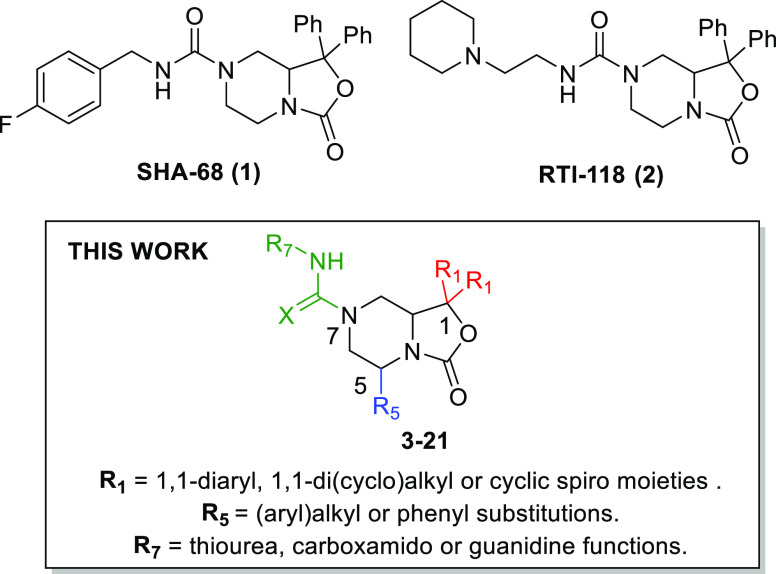
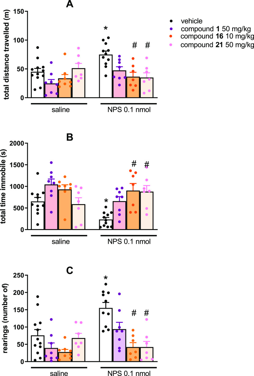
Neuropeptide S modulates important neurobiological functions including locomotion, anxiety, and drug abuse through interaction with its G protein-coupled receptor known as neuropeptide S receptor (NPSR). NPSR antagonists are potentially useful for the treatment of substance abuse disorders against which there is an urgent need for new effective therapeutic approaches. Potent NPSR antagonists in vitro have been discovered which, however, require further optimization of their in vivo pharmacological profile. This work describes a new series of NPSR antagonists of the oxazolo[3,4-a]pyrazine class. The guanidine derivative 16 exhibited nanomolar activity in vitro and 5-fold improved potency in vivo compared to SHA-68, a reference pharmacological tool in this field. Compound 16 can be considered a new tool for research studies on the translational potential of the NPSergic system. An in-depth molecular modeling investigation was also performed to gain new insights into the observed structure-activity relationships and provide an updated model of ligand/NPSR interactions.


Conflict of interest statement

The authors declare no competing financial interest.

Figures

Figure 1
Figure 1
SAR extension performed in this work around the oxazolo[3,4-a]pyrazine nucleus of known NPSR antagonists 1 and 2.
Scheme 1
Scheme 1. Synthesis of Final Compounds 3–12
Reagents and conditions: (i) BnBr, EtOH, 75 °C, 5 h; (ii) Boc2O, DMAP, tetraethylammonium, CH2Cl2, rt, 0.5 h; (iii) sec-BuLi, TMEDA, THF, −78 °C, 6 h; (iv) FmocCl, MeCN, 90 °C, 5 h; (v) DBU, THF, rt, 2 h.
Scheme 2
Scheme 2. Synthesis of Final Compounds 13–16
Reagents and conditions: (i) DBU, THF, rt, 2 h; (ii) CH2Cl2–DMF (2:1), 50 °C, 4 h; (iii) HgCl2, CH3CN, 90 °C, 24 h; (iv) BrCN,CH2Cl2, NaHCO3, H2O, 30 min at 0 °C, 24 h rt; (v) benzyl amine or 4-fluoro benzylamine, p-TsOH, DMSO, 60 °C, 18 h; (vi) K2CO3, CH3CN, 90 °C, 4 h.
Scheme 3
Scheme 3. Synthesis of Final Compounds 17–21
Reagents and conditions: (i) NaHCO3, chloroacetyl chloride, toluene, 0 °C to rt, overnight; (ii) Et3N, benzylamine, dioxane, reflux, 20 h; (iii) LiAlH4, THF, reflux, 3 h; (iv) Boc2O, THF, 0 °C to rt, 1 h; (v) benzophenone, sec-BuLi, TMEDA, THF, −78 °C, 3 h; (vi) FmocCl, MeCN, 90 °C, 5 h and then rt, 18 h; (vii) 4-fluorobenzyl isocyanate, DBU, THF, rt, 2 h.
Figure 2
Figure 2
Calcium mobilization assay performed on HEK293mNPSR cells. Concentration–response curve to NPS [panel (A)] and inhibition–response curves to 1 against the stimulatory effect of 10 nM NPS [panel (B)]. Data are mean ± SEM of at least five separate experiments made in duplicate.
Figure 3
Figure 3
Calcium mobilization assay performed on HEK293mNPSR cells. Concentration–response curve to NPS in the absence and in the presence of 100 nM of 1, 16, and 21. Data are mean ± SEM of at least five separate experiments made in duplicate.
Figure 4
Figure 4
Energy-minimized docked poses of compound 1 in BM1 and BM2 (panels A and B, respectively) in the model of NPSR constructed starting from the human neuropeptide Y Y1 receptor (hNPY1R, PDB code 5ZBH).1 and the protein are represented as violet sticks and multicolored ribbons, respectively.
Figure 5
Figure 5
Energy-minimized docked poses of compound 1 [panel (A) for BM1 and (D) for BM2], 16 [panel (B) for BM1 and (E) for BM2], and 21 [panel (C) for BM1 and (F) for BM2] in the model of NPSR constructed starting from the human neuropeptide Y Y1 receptor (hNPY1R, PDB code 5ZBH).1, 16, and 21 are represented as violet, orange, and red sticks, respectively. The protein is represented as cyan ribbons and sticks. H-bonds are represented as dashed yellow lines.
Figure 6
Figure 6
Effects of NPS, 1, 16, and 21 on mouse locomotor activity. The cumulative effects exerted on distance traveled are shown in panel (A), while the total time immobile and number of rearings over the 30 min observation period are shown in panels (B,C), respectively. Data are mean ± SEM of four separate experiments (vehicle + saline, 12 mice; 1 + saline, 8 mice; 16 + saline, 8 mice; 21 + saline, 7 mice; vehicle + NPS, 11 mice, 1 + NPS, 8 mice; 16 + NPS, 7 mice; and 21 + NPS, 7 mice). The two-way ANOVA NPS × antagonist revealed for the total distance traveled, an effect of NPS F(1,59) = 6.63 and of the interaction NPS × antagonist F(3,59) = 4.48; for the immobility time, an effect of NPS F(1,59) = 8.09 and of the interaction NPS × antagonist F(3,59) = 4.26; for the number of rearings, an effect of NPS F(1,59) = 7.35, of antagonist F(3,59) = 10.65, and of the interaction NPS × antagonist F(3,59) = 4.32. *p < 0.05 vs saline, #p < 0.05 vs vehicle according to Bonferroni’s test for multiple comparisons.

Similar articles

Cited by

References

    1. Syuji S.; Yasushi S.; Nobuyuki M.; Koji Y.. cDNA and protein sequence of a novel human and animal G protein-coupled receptors and their uses in drug screening, diagnosis, and therapeutics. WO2002031145A1, April 18, 2002.
    1. Xu Y.-L.; Reinscheid R. K.; Huitron-Resendiz S.; Clark S. D.; Wang Z.; Lin S. H.; Brucher F. A.; Zeng J.; Ly N. K.; Henriksen S. J.; de Lecea L.; Civelli O. Neuropeptide S: a neuropeptide promoting arousal and anxiolytic-like effects. Neuron 2004, 43, 487–497. 10.1016/j.neuron.2004.08.005. - DOI - PubMed
    1. Reinscheid R. K.; Xu Y.-L.; Okamura N.; Zeng J.; Chung S.; Pai R.; Wang Z.; Civelli O. Pharmacological characterization of human and murine neuropeptide S receptor variants. J. Pharmacol. Exp. Ther. 2005, 315, 1338–1345. 10.1124/jpet.105.093427. - DOI - PubMed
    1. Xu Y.-L.; Gall C. M.; Jackson V. R.; Civelli O.; Reinscheid R. K. Distribution of neuropeptide S receptor mRNA and neurochemical characteristics of neuropeptide S-expressing neurons in the rat brain. J. Comp. Neurol. 2007, 500, 84–102. 10.1002/cne.21159. - DOI - PubMed
    1. Clark S. D.; Duangdao D. M.; Schulz S.; Zhang L.; Liu X.; Xu Y.-L.; Reinscheid R. K. Anatomical characterization of the neuropeptide S system in the mouse brain by in situ hybridization and immunohistochemistry. J. Comp. Neurol. 2011, 519, 1867–1893. 10.1002/cne.22606. - DOI - PubMed

Publication types

MeSH terms

LinkOut - more resources