The metatarsaus adductus effect by the syndesmosis procedure for hallux valgus correction
- PMID: 33733823
- PMCID: PMC8009899
- DOI: 10.1302/2633-1462.23.BJO-2020-0195.R1
The metatarsaus adductus effect by the syndesmosis procedure for hallux valgus correction
Abstract
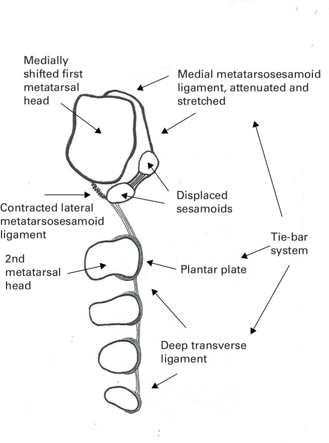
Aims: The purpose of this study is to examine the adductus impact on the second metatarsal by the nonosteotomy nonarthrodesis syndesmosis procedure for the hallux valgus deformity correction, and how it would affect the mechanical function of the forefoot in walking. For correcting the metatarsus primus varus deformity of hallux valgus feet, the syndesmosis procedure binds first metatarsal to the second metatarsal with intermetatarsal cerclage sutures.
Methods: We reviewed clinical records of a single surgical practice from its entire 2014 calendar year. In total, 71 patients (121 surgical feet) qualified for the study with a mean follow-up of 20.3 months (SD 6.2). We measured their metatarsus adductus angle with the Sgarlato's method (SMAA), and the intermetatarsal angle (IMA) and metatarsophalangeal angle (MPA) with Hardy's mid axial method. We also assessed their American Orthopaedic Foot & Ankle Society (AOFAS) clinical scale score, and photographic and pedobarographic images for clinical function results.
Results: SMAA increased from preoperative 15.9° (SD 4.9°) to 17.2° (5.0°) (p < 0.001). IMA and MPA corrected from 14.6° (SD 3.3°) and 31.9° (SD 8.0°) to 7.2° (SD 2.2°) and 18.8° (SD 6.4°) (p < 0.001), respectively. AOFAS score improved from 66.8 (SD 12.0) to 96.1 (SD 8.0) points (p < 0.001). Overall, 98% (119/121) of feet with preoperative plantar calluses had them disappeared or noticeably subsided, and 93% (113/121) of feet demonstrated pedobarographic medialization of forefoot force in walking. We reported all complications.
Conclusion: This study, for the first time, reported the previously unknown metatarsus adductus side-effect of the syndesmosis procedure. However, it did not compromise function restoration of the forefoot by evidence of our patients' plantar callus and pedobarographic findings. Level of Clinical Evidence: III Cite this article: Bone Jt Open 2021;2(3):174-180.
Keywords: Bunion; Hallux valgus; Metatarsus adductus; Metatarsus primus varus; Syndesmosis procedure.
Figures


Similar articles
-
The Syndesmosis Procedure Correction of Hallux Valgus Feet Associated With the Metatarsus Adductus Deformity.J Foot Ankle Surg. 2022 Mar-Apr;61(2):339-344. doi: 10.1053/j.jfas.2021.09.006. Epub 2021 Sep 17. J Foot Ankle Surg. 2022. PMID: 34657809
-
Radiological Analysis of the Syndesmosis Concept in Metatarsus Primus Varus and Hallux Valgus Deformities Recurrence Prevention.J Foot Ankle Surg. 2024 Mar-Apr;63(2):262-266. doi: 10.1053/j.jfas.2023.11.014. Epub 2023 Dec 5. J Foot Ankle Surg. 2024. PMID: 38056554
-
Can the Syndesmosis Procedure Prevent Metatarsus Primus Varus and Hallux Valgus Deformity Recurrence? A 5-Year Prospective Study.J Foot Ankle Surg. 2018 Mar-Apr;57(2):316-324. doi: 10.1053/j.jfas.2017.10.012. Epub 2018 Jan 12. J Foot Ankle Surg. 2018. PMID: 29336886
-
Correction of juvenile hallux valgus deformity associated with metatarsus primus adductus using epiphysiodesis technique.Clin Podiatr Med Surg. 1987 Jan;4(1):63-74. Clin Podiatr Med Surg. 1987. PMID: 2949818 Review.
-
Hallux valgus and metatarsus adductus: the surgical dilemma.Clin Podiatr Med Surg. 1989 Jan;6(1):103-13. Clin Podiatr Med Surg. 1989. PMID: 2653602 Review.
References
-
- Truslow W. Metatarsus Primus varus or hallux valgus. JBJS. 1925;7A(1):98–108.
-
- Kimura T, Kubota M, Taguchi T, Suzuki N, Hattori A, Marumo K. Evaluation of First-Ray mobility in patients with hallux valgus using weight-bearing CT and a 3-D analysis system: a comparison with normal feet. J Bone Joint Surg Am. 2017;99-A(3):247–255. - PubMed
-
- Shibuya N, Roukis TS, Jupiter DC. Mobility of the first ray in patients with or without hallux valgus deformity: systematic review and meta-analysis. J Foot Ankle Surg. 2017;56(5):1070–1075. - PubMed
-
- Pinney S, Song K, Chou L. Surgical treatment of mild hallux valgus deformity: the state of practice among academic foot and ankle surgeons. Foot Ankle Int. 2006;27(11):970–973. - PubMed
-
- Pinney SJ, Song KR, Chou LB. Surgical treatment of severe hallux valgus: the state of practice among academic foot and ankle surgeons. Foot Ankle Int.. 2006;27(12):1024–1029. - PubMed
LinkOut - more resources
Full Text Sources
Other Literature Sources