. 2021 May;35(3):1455-1465.
doi: 10.1111/jvim.16085. Epub 2021 Mar 18.

A hypomyelinating leukodystrophy in German Shepherd dogs

Affiliations

A hypomyelinating leukodystrophy in German Shepherd dogs

Pia R Quitt et al. J Vet Intern Med. 2021 May.

Abstract

Background: Shaking puppy syndrome is commonly attributed to abnormal myelination of the central nervous system.

Hypothesis/objectives: To report the long-term clinical course and the imaging characteristics of hypomyelinating leukodystrophy in German Shepherd dogs.

Animals and methods: Three related litters with 11 affected dogs.

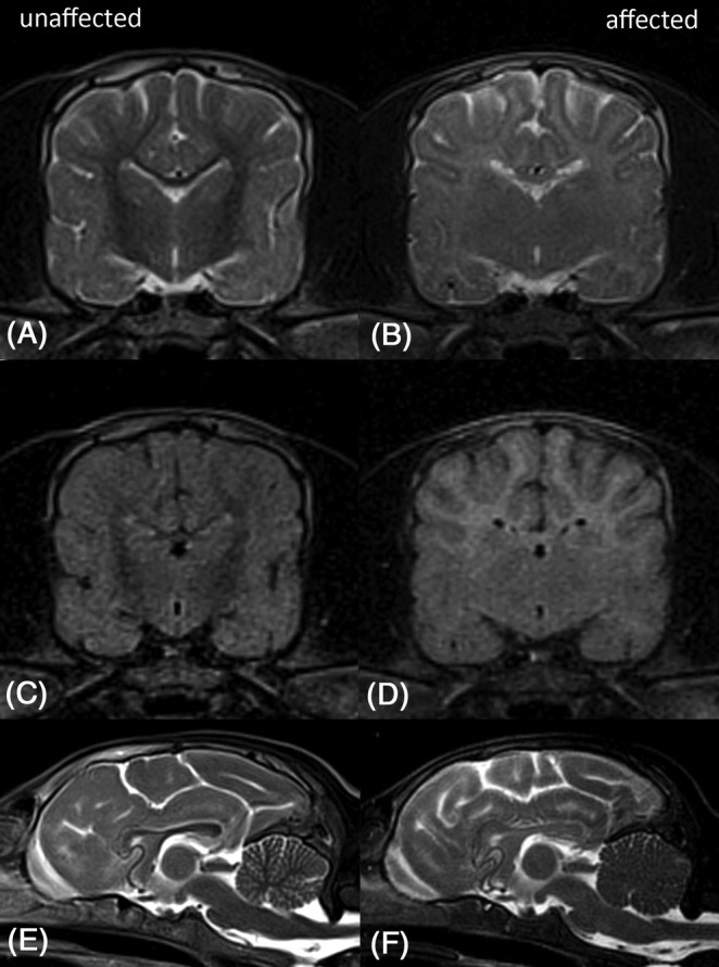
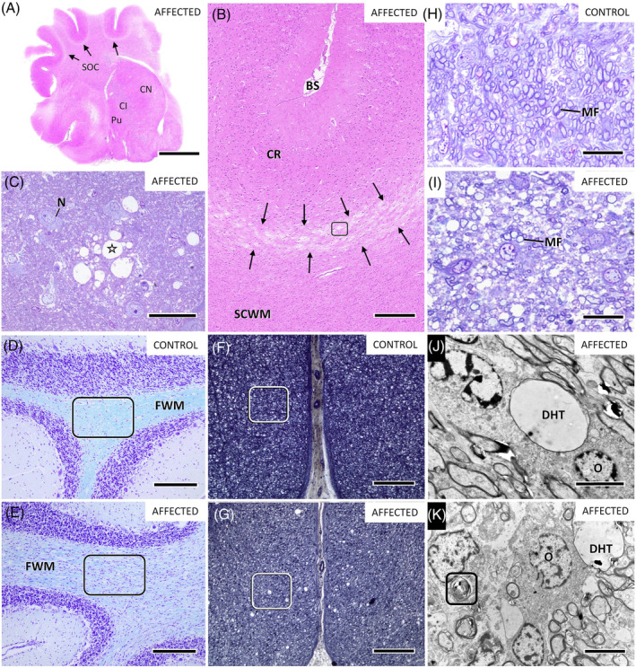
Results: The 11 affected dogs experienced coarse, side-to-side tremors of the head and trunk, which interfered with normal goal-oriented movements and disappeared at rest. Signs were noticed shortly after birth. Nine dogs were euthanized, 3 dogs underwent pathological examination, and 2 littermates were raised by their breeder. Tremors improved gradually until 6 to 7 months of age. Adult dogs walked with severe residual pelvic limb ataxia. One dog developed epilepsy with tonic-clonic seizures at 15 months of age. Conventional magnetic resonance imaging (MRI) disclosed homogenous hyperintense signal of the entire subcortical white matter in 3 affected 7-week-old dogs and a hypointense signal in a presumably unaffected littermate. Subcortical white matter appeared isointense to gray matter at 15 and 27 weeks of age on repeated MRI. Abnormal white matter signal with failure to display normal gray-white matter contrast persisted into adulthood. Cerebellar arbor vitae was not visible at any time point. Clinical signs, MRI findings, and pathological examinations were indicative of a hypomyelinating leukodystrophy. All parents of the affected litters shared a common ancestor and relatedness of the puppies suggested an autosomal recessive mode of inheritance.

Conclusion: We describe a novel hypomyelinating leukodystrophy in German Shepherd dogs with a suspected inherited origin.

Keywords: animal model; brain maturation; development; dysmyelination; genetic; hypomyelination; inherited; leukoencephalopathy; seizures; tremor; white matter.


Conflict of interest statement

Authors declare no conflict of interest.

Figures

FIGURE 1
FIGURE 1
Brain MRI at 7 weeks of age. The figure displays MRI from the unaffected and 1 affected littermate (dog 2) at 7 weeks of age. The upper row shows T2W transverse images (A, B) at the level of the interthalamic adhesion, the middle row shows corresponding transverse FLAIR images (C, D), and the lower row shows T2W midsagittal images (E, F). Subcortical white matter appears hypointense compared to gray matter in the unaffected littermate. In contrast, subcortical white matter appears homogeneously hyperintense in the affected littermate. Corpus callosum appears hypointense to gray matter in the unaffected littermate (E) and isointense in the affected dog (F). The affected dog shows poor differentiation between cerebellar gray and white matter and few hyperintense patches in the brainstem on midsagittal T2W images (F). FLAIR, fluid attenuation inversion recovery; MRI, magnetic resonance imaging
FIGURE 2
FIGURE 2
T2W dorsal brain MRI at 7 weeks of age. The figure displays dorsal T2W images of the brain from the unaffected dog and 1 affected dog at 7 weeks of age. Subcortical white matter appears homogenously hypointense to gray matter in the unaffected littermate (A) and hyperintense in the affected dog (B). MRI, magnetic resonance imaging
FIGURE 3
FIGURE 3
Follow‐up brain MRI at 15 weeks of age. The figure displays MRI of the brain from the affected dog 3 at 7 and 15 weeks of age. The upper row shows T2W transverse images at the level of the interthalamic adhesion (A, B), the middle row shows corresponding transverse FLAIR images (C, D), and the lower row shows T2W midsagittal images (E, F). Subcortical white matter appears hyperintense to gray matter at 7 weeks and isointense at 15 weeks of age on T2W and FLAIR images. Thus the affected dog failed to display the normal transition phase to white matter hypointensity, which occurs in healthy Beagles at 6 weeks (T2W) and 8 to 36 weeks of age (FLAIR). 26 FLAIR sequence failed in this dog at 7 weeks of age presumably due to artifact (C). 30 FLAIR, fluid attenuation inversion recovery; MRI, magnetic resonance imaging
FIGURE 4
FIGURE 4
Follow‐up brain MRI at 27 and 69 weeks of age. The figure displays MRI of the brain from the affected dog 3 at 27 and 69 weeks of age. The upper row shows T2W transverse images at the level of the interthalamic adhesion (A, B), the middle row shows corresponding transverse FLAIR images (C, D), and the lower row shows T2W midsagittal images (E, F). Subcortical white matter appears isointense to gray matter at 27 weeks of age on T2W and FLAIR images (A, C). In the adult dog, at 69 weeks of age, the subcortical (B, D) and cerebellar white matter still appeared isointense to mildly hypointense without normal gray‐white matter contrast (F). The cerebellar arbor vitae is not discernible (E, F). On midsagittal images, the corpus callosum appears more hypointense at 27 and 69 weeks of age (E, F). FLAIR, fluid attenuation inversion recovery; MRI, magnetic resonance imaging
FIGURE 5
FIGURE 5
Brain MRI from one adult affected dog and a control dog. The figure displays T2W MRI from one affected dog (A, C) at 69 weeks of age (dog 3), and a control dog (B, D; 4‐month‐old). The upper row shows T2W midsagittal images at the level of the interthalamic adhesion (A, B) and the lower row shows the corresponding transverse images (C, D). The affected adult dog fails to display normal gray‐white‐matter contrast in the cerebrum (C) and cerebellum (A). The cerebellar arbor vitae is not visible on midsagittal images (A). The entire subcortical and cerebellar white matter appears isointense to mildly hypointense compared to gray matter. The control dog shows prominent hypointensity of the subcortical white matter (D) and a clearly visible cerebellar arbor vitae (B). MRI, magnetic resonance imaging
FIGURE 6
FIGURE 6
Representative neuropathological findings in frontal cortex (A‐C), subcortical white matter (B, I, K), capsula interna (J), cerebellum (E), and thoracic spinal cord (G) of an affected dog (D, 7 weeks) and an age‐matched control (D, F, H). In the frontal cortex, at subgross magnification, a pale line (A: arrows) signifies edema and spongiosis (B: black arrows) of inner cortical laminae and white‐gray matter interface. Vacuolation of cortical neuropil (B: frame, C: asterisk) is a consequence to myelin splitting (not shown). Subcortical white matter (B: SCWM) including semioval center (A: SOC) stains abnormally pale and shows widespread neuroglial hypercellularity (B). Hypomyelination of central white matter becomes more evident using myelin specific dyes LFB (D, E) and WSS (F, G). Attenuation of myelin staining is demonstrated in foliary white matter (FWM) of cerebellum and in long spinal tracts of the affected dog (E, G) compared to the age‐matched control (D, F); framed areas indicate the neuroglial white matter hypercellularity (E) and a mild white matter spongiosis of ventral funiculus (G). On semithin sections (H, I), myelinated fibers (MFs) in the affected dog (I) are sparse and present with variable calibers and degrees of myelination when compared to the control dog (H). Affected white matter shows microvacuolation (I) and contains numerous poorly myelinated fibers, dilated hypomyelinated myelin tubes (J, K: DHT) and degenerate myelin figures (K: frame). Oligodendrocytes appear hypertrophic (J, K: O) and show increased number and density (I). Stains and contrast agents: (A, B): Hematoxylin‐Eosin (HE); (C, H, I): azure II methylene blue‐safranin O (ASO); (D, E): Luxol fast blue‐Cresyl echt violet (LFB); (F, G): Woelcke‐Spielmeyer‐Schröder stain (WSS); (J, K): uranyl acetate and lead citrate. Anatomical and histological labels: BS, base of sulcus; CI, capsula interna; CN, caudate nucleus; CR, cortical ribbon; FWM, foliary white matter; MF, myelinated fiber; N, neuron; O, oligodendrocyte; Pu, putamen; SCWM, subcortical white matter; SOC, semioval center. Magnification scale bars: (A): 0.5 cm; (B): 370 μm; (C): 25 μm; (D, E): 200 μm; (F, G): 100 μm; (H, I): 12.5 μm; (J): 2500 nm; (K): 3000 nm
FIGURE 7
FIGURE 7
Pedigree of German shepherd dogs including two closely related German and one Finnish litter with multiple affected dogs. The parents of the affected litters share a common ancestor (red circle). The pedigree is consistent with an autosomal recessive mode of inheritance

References

    1. Vanderver A, Prust M, Tonduti D, et al. Case definition and classification of leukodystrophies and leukoencephalopathies. Mol Genet Metab. 2015;114:494‐500. 10.1016/j.ymgme.2015.01.006. - DOI - PMC - PubMed
    1. Van der Knaap MS, Bugiani M. Leukodystrophies: a proposed classification system based on pathological changes and pathogenetic mechanisms. Acta Neuropathol. 2017;134:351‐382. 10.1007/s00401-017-1739-1. - DOI - PMC - PubMed
    1. Duncan ID. Abnormalities of myelination of the central nervous system associated with congenital tremor. J Vet Intern Med. 1987;1:10‐23. - PubMed
    1. Cummings JF, Summers BA, de Lahunta A, Lawson C. Tremors in Samoyed pups with oligodendrocyte deficiencies and hypomyelination. Acta Neuropathol. 1986;71:267‐277. - PubMed
    1. Palmer AC, Blakemore WF, Wallace ME, Wilkes MK, Herrtage ME, Matic SE. Recognition of 'trembler', a hypomyelinating condition in the Bernese mountain dog. Vet Rec. 1987;120:609‐612. - PubMed