. 2021 Mar 18;17(3):e1009324.
doi: 10.1371/journal.ppat.1009324. eCollection 2021 Mar.

Structural basis of Blastomyces Endoglucanase-2 adjuvancy in anti-fungal and -viral immunity

Affiliations

Structural basis of Blastomyces Endoglucanase-2 adjuvancy in anti-fungal and -viral immunity

Lucas Dos Santos Dias et al. PLoS Pathog. .

Abstract

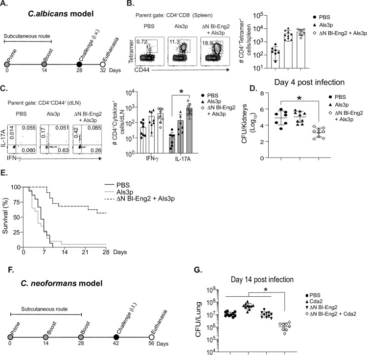
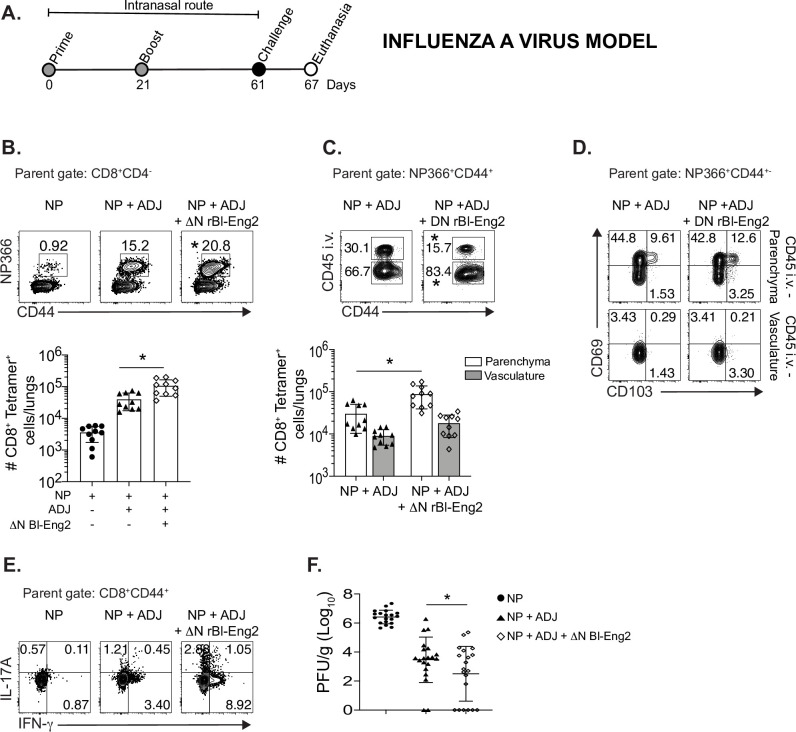
The development of safe subunit vaccines requires adjuvants that augment immunogenicity of non-replicating protein-based antigens. Current vaccines against infectious diseases preferentially induce protective antibodies driven by adjuvants such as alum. However, the contribution of antibody to host defense is limited for certain classes of infectious diseases such as fungi, whereas animal studies and clinical observations implicate cellular immunity as an essential component of the resolution of fungal pathogens. Here, we decipher the structural bases of a newly identified glycoprotein ligand of Dectin-2 with potent adjuvancy, Blastomyces endoglucanase-2 (Bl-Eng2). We also pinpoint the developmental steps of antigen-specific CD4+ and CD8+ T responses augmented by Bl-Eng2 including expansion, differentiation and tissue residency. Dectin-2 ligation led to successful systemic and mucosal vaccination against invasive fungal infection and Influenza A infection, respectively. O-linked glycans on Bl-Eng2 applied at the skin and respiratory mucosa greatly augment vaccine subunit- induced protective immunity against lethal influenza and fungal pulmonary challenge.


Conflict of interest statement

The authors have declared that no competing interests exist.

Figures

Fig 1
Fig 1. Dectin-2 recognizes mannosylation but not the protein backbone of Bl-Eng2.
A) Recombinant (r) Bl-Eng2 expressed in E. coli (the higher molecular band in the red rectangular box was confirmed to be Bl-Eng2 by western blot) or P. pastoris and analyzed by SDS-PAGE. B) B3Z reporter cells expressing FcγR plus Dectin-2 or FcγR (B3Z) alone (negative control) were incubated with rBl-Eng2 from P. pastoris (30 ng) or E. coli (30-240ng) and reporter activity measured. C) rBl-Eng2 from P. pastoris was deglycosylated with trifluormethanesulfonic acid (TFMS) and analyzed by SDS-PAGE. Colloidal Coomassie stain was used to evaluate the molecular size and Pro-Q Emerald 300 to evaluate glycosylation. D) Deglycosylated and proteinase K-treated rBl-Eng2 from P. pastoris was analyzed by Dectin-2 reporter assay. E) rBl-Eng2 from P. pastoris was tested for Dectin-2 reporter activity in the presence of soluble glucose, galactose or mannose. F) NFAT-GFP reporter cells expressing wild-type Dectin-2 or a mutant CRD that doesn’t recognize mannose were stimulated with rBl-Eng2 and analyzed for GFP expression by flow cytometry. The data are representative of three independent experiments.
Fig 2
Fig 2. Glycosylation profiling of Bl-Eng2.
(A) Glycan modificatons of Bl-Eng2 identified included N-linked (indicated by square) and O-linked glycans (indicated by circle) with (shown as red with P) and without phosphorylation. (B) Glycosylation sites and glycoform annotation of rBl-Eng2. The O-linked mannose residues at the N-terminus, including the catalytic GH16 domain and surrounding sequence, are short and range from 1 to 3 mannoses. The number in the circle indicates the number of mannose residues. Green circle means mannose, red circle means phosphorylated mannose, with the number in red circle indicating the position of mannose being phosphorylated. As shown, the O-linked glycans at the C-terminus are generally longer and have size ranging between 2 and 21 mannose residues. Glycosylation of the Serine/Threonine rich region was not profiled due to lack of enzyme cleavage sites. (C) Annotated electron transfer/higher-energy collision dissociation (EThcD) tandem mass spectrum of a representative O-linked glycopeptide (QKLISHex(7)phosphoEE) of rBl-Eng2, Hex = Mannose. Singly charged Y-ions (peptide plus glycan remnant) are annotated along the top. Blue asterisks (*) represent the deamination peaks. A series of sequence-specific b (labeled in blue) and y (labeled in red) type of fragment ions enable derivation of amino acid sequence of the peptide. A series of sequential loss of mannose peaks (-162 Da, indicated by green circles between two dashed lines above the blue double arrowhead) suggests a total of 7 mannose residues modified at the serine residue. Two adjacent peaks with identical number of mannose residues with 80 Da mass difference (shown as–P above the yellow double arrowhead) suggest the loss of phosphorylation, which modifies the 2nd mannose residue attached to the serine residue of the O-glycopeptide. The oxonium ions generated from glycan cleavage by HCD suggest the presence of various glycan moieties, annotated in green, in the low m/z region. (D) Glycan linkage information revealed by GCMS analysis. 1,2-linked mannose and terminal mannose are the major linkages of the glycans, with trace amounts of 1,4-linked Mannose, 1,6-linked mannose, 2,3-linked mannose and 2,6-linked mannose. Relative percentages of these glycans are shown on the right side of the panel.
Fig 3
Fig 3. The C-terminus of Bl-Eng2 stimulates Dectin-2 reporter activity.
A) rBl-Eng2 lacking the C-terminus or the N-terminus was expressed in P. pastoris. B) Full length and truncated proteins were purified and analyzed by SDS-PAGE. C+D) All three proteins were tested in the reporter assay by titrating mass (C) or molar amounts (D). E+F) BMDCs from wild type and Dectin-2-/- mice were stimulated with rBl-Eng2 constructs and IL-6 (E) and IL-1β (F) measured in cell culture supernatants by ELISA. Stimulation with B. dermatitidis yeast served as a positive control. *p<0.5 vs. full length Bl-Eng2. The data are representative of three independent experiments.
Fig 4
Fig 4. The C-terminus of Bl-Eng2 (ΔN rBl-Eng2) augments T cell expansion, tissue residency, Th1 and Th17 cell differentiation and resistance upon vaccination against B. dermatitidis infection.
Two antigens were used to test adjuvant activity: calnexin (CNX) (A+B), and Bl-Eng2 peptide (C-F). (A+B) Mice received adoptively transferred 1807 T cells prior to vaccination and were subcutaneously vaccinated with 240 ng calnexin peptide and 10μg ΔN rBl-Eng2 (as adjuvant). Two weeks after the boost, mice were challenged with virulent B. dermatitidis yeast. At day 4 post-infection, the frequency (A) and number (B) of lung 1807 cells (CD90.1+) producing IL-17 and IFN-γ were analyzed by flow cytometry. C-F) Mice were vaccinated with Bl-Eng2 peptide and ΔN rBl-Eng2 as above and challenged with B. dermatitidis. At day 4 post-infection, the frequency (C) and number (D) of total tetramer+ T cells in the lungs (top row) are shown, including those in the parenchyma and vasculature (middle row), and those producting IL-17 and IFNγ (bottom row) as analyzed by flow cytometry. Data are from a representative experiment (n = 5 mice/group) of two performed. *p<0.5 vs. the Bl-Eng2 or calnexin peptide alone group. E) Lung CFU are displayed as the geometric mean with standard deviation. *p<0.05 vs. Bl-Eng2 peptide group. F) Percent body weight change was calculated by weighing the mice at the time of challenge and euthanasia (two weeks post infection). N = 10 mice/group. *p<0.05 vs. Bl-Eng2 peptide group.
Fig 5
Fig 5. Adjuvant activity of the C-terminus of Bl-Eng2 (ΔN rBl-Eng2) is glycosylation- and Dectin-2 dependent.
Wild type mice were vaccinated with deglycosylated ΔN rBl-Eng2 and Dectin-2-/- mice received untreated ΔN rBl-Eng2. Percentage (A) and number (B) of tetramer+ lung T cells were analyzed 4 days post-infection. Percentage (C) and number (D) of IL-17A and IFN-γ producing Bl-Eng2-specific CD4+ T cells. Data represent an average of two independent experiments (n = 10 mice/group). *p<0.05 for WT vs KO vaccine groups. E) Lung CFU are displayed as geometric mean with standard deviation. *p<0.05 for WT vs KO vaccine groups. F) IFNγ and IL-17A from cell culture supernatants of Bl-Eng2 peptide-stimulated splenocytes as measured by ELISA. *p<0.05 for vaccinated WT vs. KO groups.
Fig 6
Fig 6. Vaccination with ΔN rBl-Eng2 augments protective immunity against systemic candidiasis and pulmonary cryptococcosis.
A) Mice were subcutaneously vaccinated twice, two weeks apart with ΔN rBl-Eng2 and Als3 peptide and challenged with C. albicans strain ATCC SC5314 (intravenously). B) The frequency and number of Als3-specific CD4+ T cells in the spleen at day 4 post-challenge after tetramer pull down. C) The percentage and number of IL-17A and IFN-γ producing Als3-specific CD4+ T cells in the draining lymph nodes at day 4 post-infection. D) C. albicans CFU in the kidneys at day 4 post-challenge and E) survival curve. F) Mice were SC vaccinated three times, two weeks apart and challenged with C. neoformans var grubii strain KN99 (intracheally) two weeks after the last boost. G) C. neoformans lung CFU at day 14 post-infection. Data are the average of two indendepent experiments (n = 10 mice/group for cellular analysis and CFU. n = 20 mice/group for survival). *p<0.05 for ΔN rBl-Eng2 + Als3 or Cda2 vs PBS.
Fig 7
Fig 7. The C-terminus of Bl-Eng2 (ΔN rBl-Eng2) serves as an adjuvant, augmenting influenza specific CD8+ T cell expansion, tissue residency, Th17 and Tc17 cell differentiation and resistance to IAV infection.
A) Mice were intranasally vaccinated with nucleoprotein (NP), adjuplex and/or ΔN rBl-Eng2 protein and challenged with influenza A virus (IAV) (strain PR8). B) CD8+ NP366-specific tetramer+ T cells were enumerated in the lungs day 6 post infection. C) Anti-CD45 mAb was injected i.v 3 minutes before euthanizing mice to distinguish and enumerate parenchymal (CD45-) from vasculature associated lung cells (CD45+). D) CD69 and CD103 expression by parenchymal (CD45-) vs. vascular (CD45+) NP366-specific CD8+ T cells. E) IL-17A and IFN-γ production by peptide-stimulated lung CD8+ T cells. F) Viral load in the left lung was assessed by plaque assay. Data are the average of two independent experiments performed (n = 20 mice/group). Male and female cohorts of mice were used for these experiments and no gender difference was observed. *p<0.05 for adjuplex + NP vs Adjuplex + NP + ΔN rBl-Eng2.

Similar articles

Cited by

References

    1. Ellebedy AH, Webby RJ. Influenza vaccines. Vaccine. 2009;27 Suppl 4:D65–8. Epub 2009/10/20. S0264-410X(09)01214-6 [pii] 10.1016/j.vaccine.2009.08.038 - DOI - PMC - PubMed
    1. Schotsaert M, Saelens X, Leroux-Roels G. Influenza vaccines: T-cell responses deserve more attention. Expert Rev Vaccines. 2012;11(8):949–62. Epub 2012/09/26. 10.1586/erv.12.71 . - DOI - PubMed
    1. Blackburne BP, Hay AJ, Goldstein RA. Changing selective pressure during antigenic changes in human influenza H3. PLoS Pathog. 2008;4(5):e1000058. Epub 2008/05/03. 10.1371/journal.ppat.1000058 - DOI - PMC - PubMed
    1. Tricco AC, Chit A, Soobiah C, Hallett D, Meier G, Chen MH, et al.. Comparing influenza vaccine efficacy against mismatched and matched strains: a systematic review and meta-analysis. BMC Med. 2013;11:153. Epub 2013/06/27. 1741-7015-11-153 [pii] 10.1186/1741-7015-11-153 - DOI - PMC - PubMed
    1. Yap KL, Ada GL, McKenzie IF. Transfer of specific cytotoxic T lymphocytes protects mice inoculated with influenza virus. Nature. 1978;273(5659):238–9. Epub 1978/05/18. 10.1038/273238a0 . - DOI - PubMed

Publication types

MeSH terms