. 2021 Mar 18;17(3):e1009403.
doi: 10.1371/journal.ppat.1009403. eCollection 2021 Mar.

A swine arterivirus deubiquitinase stabilizes two major envelope proteins and promotes production of viral progeny

Affiliations

A swine arterivirus deubiquitinase stabilizes two major envelope proteins and promotes production of viral progeny

Rui Guo et al. PLoS Pathog. .

Abstract

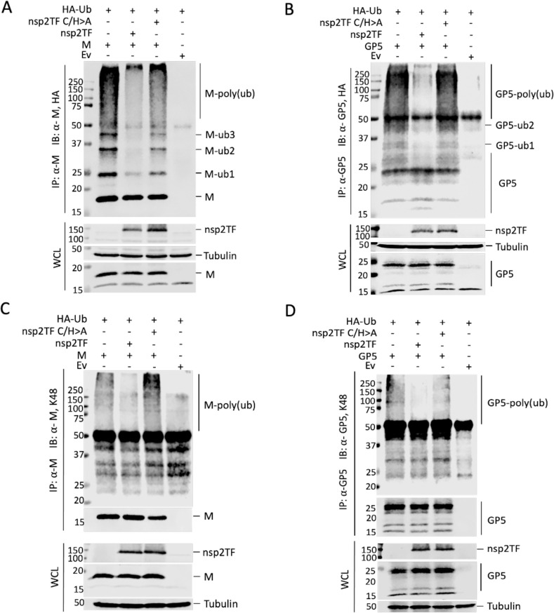
Arteriviruses are enveloped positive-strand RNA viruses that assemble and egress using the host cell's exocytic pathway. In previous studies, we demonstrated that most arteriviruses use a unique -2 ribosomal frameshifting mechanism to produce a C-terminally modified variant of their nonstructural protein 2 (nsp2). Like full-length nsp2, the N-terminal domain of this frameshift product, nsp2TF, contains a papain-like protease (PLP2) that has deubiquitinating (DUB) activity, in addition to its role in proteolytic processing of replicase polyproteins. In cells infected with porcine reproductive and respiratory syndrome virus (PRRSV), nsp2TF localizes to compartments of the exocytic pathway, specifically endoplasmic reticulum-Golgi intermediate compartment (ERGIC) and Golgi complex. Here, we show that nsp2TF interacts with the two major viral envelope proteins, the GP5 glycoprotein and membrane (M) protein, which drive the key process of arterivirus assembly and budding. The PRRSV GP5 and M proteins were found to be poly-ubiquitinated, both in an expression system and in cells infected with an nsp2TF-deficient mutant virus. In contrast, ubiquitinated GP5 and M proteins did not accumulate in cells infected with the wild-type, nsp2TF-expressing virus. Further analysis implicated the DUB activity of the nsp2TF PLP2 domain in deconjugation of ubiquitin from GP5/M proteins, thus antagonizing proteasomal degradation of these key viral structural proteins. Our findings suggest that nsp2TF is targeted to the exocytic pathway to reduce proteasome-driven turnover of GP5/M proteins, thus promoting the formation of GP5-M dimers that are critical for arterivirus assembly.


Conflict of interest statement

The authors have declared that no competing interests exist.

Figures

Fig 1
Fig 1. Co-localization analysis of nsp2TF with other PRRSV nonstructural and structural proteins.
MARC-145 cells were infected with PRRSV SD95-21 at an moi of 0.1. The cells were fixed at 8, 10 or 12 hpi, permeabilized, and immunostained for nsp1α, nsp1β, nsp4, nsp7, nsp8, GP3, GP4, GP5, M and N proteins together with nsp2TF or nsp2. (A and B) Double labeling for GP5 (green) and nsp2TF (A) or nsp2 (B) in cells fixed at 10 hpi. (C and D) Double labeling for M protein (green) and nsp2TF (C) or nsp2 (D) in cells fixed at 10 hpi. (E) Double labeling for GP5 (green) and M protein (red) in cells fixed at 10 hpi. (F-H) Analysis of co-localization between nsp1α, nsp1β, nsp4, nsp7, nsp8, GP3, GP4, GP5, M or N protein and nsp2TF or nsp2 in PRRSV-infected MARC-145 cells fixed at 8 (F), 10 (G), or 12 (H) hpi. Nsp2TF or nsp2 foci were chosen as regions of interest for Pearson’s correlation coefficient analysis; 10 foci were randomly selected for each sample (for details, see Materials and Methods). Scale bars, 10μm. ***p < 0.001, *p<0.05. ns, statistically not significant.
Fig 2
Fig 2. Interaction of nsp2TF with GP5 and M protein.
(A-C) MARC-145 cells were mock-infected or infected with PRRSV. (A) Cell lysates were collected at 8, 10 and 12 hpi and subjected to IP. Lysates were immunoprecipitated with anti-nsp2TF pAb or anti-nsp2 pAb. Western blot analysis was conducted with the whole cell lysate or immunoprecipitated proteins. Membranes were probed with anti-nsp2TF and anti-nsp2 pAbs, or anti-M and anti-GP5 mAbs. (B-C) Cell lysates were collected at 12 hpi and subjected to co-IP and western blot analysis. Lysates were immunoprecipitated with anti-M pAb (B) or anti-GP5 pAb (C) and membranes were probed with anti-nsp2TF pAb, anti-PLP2 mAb, or anti-M and anti-GP5 mAbs. (D-G) HEK-293T cells were co-transfected with a plasmid expressing nsp2TF and a plasmid expressing M protein (D-E) or GP5 (F-G). Cell lysates were collected at 48 h post-transfection and subjected to co-IP using a specific pAb against each individual protein as indicated in each panel. Membranes were probed with anti-nsp2TF pAb, anti-M or anti-GP5 mAb. Anti-tubulin mAb was used as a control. IP: immunoprecipitation; IB: immunoblotting; WCL: Whole cell lysates; Ev: empty vector.
Fig 3
Fig 3. Co-localization analysis between nsp2TF and organelle-specific cellular marker proteins in the secretory pathway.
MARC-145 cells were infected with PRRSV SD95-21. At 10 hpi, cells were fixed, permeabilized and immunostained for nsp2TF (A), GP5 (B), M protein (C) and nsp2 (D). To label specific cellular compartments, the cells were co-stained with antibodies specific for ERGIC marker proteins ERGIC-53, COPA, COPB, or Golgin 97. Scale bars, 10 μm. (E) The level of co-localization between ERGIC makers and nsp2, nsp2TF, GP5 or M protein quantified by Pearson’s correlation coefficient using the Coloc2 module of the ImageJ software package. Mean (± SD) values from three replicates are shown. ***p < 0.001.
Fig 4
Fig 4. Polyubiquitination of PRRSV GP5 and M protein.
HEK-293T cells were co-transfected with plasmids expressing HA-Ub, GP5 and/or M protein. At 42 h post transfection, proteasome inhibitor MG132 was added. At 48 h post transfection, cell lysates were collected for IP analysis. (A and C) Lysates were immunoprecipitated with anti-M pAb. (B and D) Lysates were immunoprecipitated with anti-GP5 pAb. Western blot analysis was conducted with the whole cell lysate or immunoprecipitated proteins using the mAb against HA, antibodies specifically recognize K27-, K48- or K63-linked polyubiquitin chains, or a specific Ab to each viral protein as labeled for each panel. IP: immunoprecipitation; IB: immunoblotting; WCL: whole cell lysates; Ev: empty vector.
Fig 5
Fig 5. Nsp2TF antagonizes polyubiquitination of GP5 and M protein.
HEK-293T cells were co-transfected with plasmids expressing HA-Ub, M protein (A and C) or GP5 (B and D) and a plasmid expressing nsp2TF or the PLP2 catalytic sites mutant (nsp2TF-C/H>A). Cell lysates were collected at 48 h post transfection. IP was performed with anti-M pAb (A and C) or anti-GP5 pAb (B and D) and the membrane was probed with anti-HA mAb (A and B), a pAb recognizing anti-K48-linked polyubiquitin (C and D), or a viral protein-specific antibody. IP: immunoprecipitation; IB: immunoblotting; WCL: whole cell lysates; Ev: empty vector.
Fig 6
Fig 6. Construction and characterization of PRRSV mutant vKTF.
(A) Schematic diagram of the engineered nsp2TF truncation. The stop codon introduced in the construct is labeled with an asterisk. The truncated region is shaded with gray. HVR, hypervariable region; nsp2 C-term: nsp2 C-terminal domain that is distinct from that of nsp2TF. (B-C) MARC-145 cells were infected with WT virus or its mutant vKTF. Cell lysates were harvested every 12 hpi. (B) Growth kinetics comparison of WT virus and vKTF mutant. Each data point represents the mean of three replicates. Virus titers are presented as numbers of fluorescent-focus units (FFU) per milliliter (ml). ****p < 0.0001. (C) Cell lysate harvested at 36 hpi was subjected to Western blot analysis using anti-PLP2 mAb that recognizes the common N-terminal PLP2 domain present in all three nsp2 variants (top panel), the TF-specific pAb (middle panel), or mAbs specific for M and GP5 proteins (bottom panel). Anti-tubulin mAb was used as a control.
Fig 7
Fig 7. Nsp2TF deubiquitinating GP5 and M protein in the context of PRRSV-infected cells.
(A-B) HEK-293T cells were co-transfected with a plasmid expressing HA-Ub and a PRRSV full-length infectious cDNA clone of WT virus or its mutant vKTF. The cell lysates were collected at 48 h post-transfection. IP was performed with anti-M pAb (A) or anti-GP5 pAb (B). Immunoprecipitated proteins were separated by SDS-PAGE and analyzed by Western blot using anti-HA mAb (A-B). Western blot analysis was also conducted with pre-IP whole cell lysates using M-specific mAb (A) or GP5 specific mAb (B). Tubulin was detected as a loading control. IP: immunoprecipitation; IB: immunoblotting; WCL: whole cell lysates.
Fig 8
Fig 8. Nsp2TF protects GP5 and M protein from UPP degradation.
(A-P) HEK-293T cells were transfected with a plasmid expressing GP5 or M protein, or co-transfected with plasmids expressing the nsp2TF (or its mutant nsp2TF C/H>A) and GP5 or M protein. At 24 h post-transfection, transfected cells were treated with CHX alone or in combination with the proteasome inhibitor MG132. For each transfection condition, cells were collected every 2 h after the drug treatment. The whole cell lysates were used for Western blot analysis. Nsp2TF, GP5 and M protein expression were detected with anti-nsp2TF pAb, anti-M pAb and anti-GP5 mAb. β-tubulin was included as a loading control. (B, D, F, H) Relative protein expression levels of M protein under the corresponding experimental condition in the left panels A, C, E, G, respectively. (J, L, N, P) Relative protein expression levels of GP5 under the corresponding experimental condition in the left panels I, K, M, O, respectively. (Q, S, U, W) MARC-145 cells were infected with WT virus (panels Q and S) or mutant vKTF (panels U and W). At 12 hpi, the cells were treated with CHX alone or together with MG132. Cell lysates were collected every 2 h. The whole cell lysates were used for Western blot analysis. Viral protein expression was detected with a specific antibody as described above. (R, T, V, X) The relative expression levels of GP5 and M protein under the experimental conditions in panels Q, S, U, W, respectively. Each graph for relative protein expression level was built based on two independent experiments. ****p < 0.0001, ***p < 0.001, **p < 0.01, *p < 0.05. ns, statistically not significant.

References

    1. Snijder EJ, Kikkert M, Fang Y. Arterivirus molecular biology and pathogenesis. The Journal of general virology. 2013;94(Pt 10):2141–63. Epub 2013/08/14. 10.1099/vir.0.056341-0 - DOI - PubMed
    1. Kuhn JH, Lauck M, Bailey AL, Shchetinin AM, Vishnevskaya TV, Bao Y, et al.. Reorganization and expansion of the nidoviral family Arteriviridae. Arch Virol. 2016;161(3):755–68. Epub 2015/11/27. 10.1007/s00705-015-2672-z [pii]. - DOI - PMC - PubMed
    1. Gulyaeva A, Dunowska M, Hoogendoorn E, Giles J, Samborskiy D, Gorbalenya AE. Domain Organization and Evolution of the Highly Divergent 5’ Coding Region of Genomes of Arteriviruses, Including the Novel Possum Nidovirus. J Virol. 2017;91(6). Epub 2017/01/06. e02096-16 [pii] 10.1128/JVI.02096-16 - DOI - PMC - PubMed
    1. Dokland T. The structural biology of PRRSV. Virus Res. 2010;154(1–2):86–97. Epub 2010/08/10. 10.1016/j.virusres.2010.07.029 - DOI - PMC - PubMed
    1. de Vries AA, Chirnside ED, Horzinek MC, Rottier PJ. Structural proteins of equine arteritis virus. J Virol. 1992;66(11):6294–303. 10.1128/JVI.66.11.6294-6303.1992 - DOI - PMC - PubMed

Publication types

MeSH terms