Review
. 2021 Mar 18;17(3):e1009342.
doi: 10.1371/journal.ppat.1009342. eCollection 2021 Mar.

The granuloma in cryptococcal disease

Affiliations
Review

The granuloma in cryptococcal disease

Laura C Ristow et al. PLoS Pathog. .

Abstract

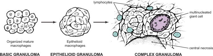
Although we have recognized cryptococcosis as a disease entity for well over 100 years, there are many details about its pathogenesis which remain unknown. A major barrier to better understanding is the very broad range of clinical and pathological forms cryptococcal infections can take. One such form has been historically called the cryptococcal granuloma, or the cryptococcoma. These words have been used to describe essentially any mass lesion associated with infection, due to their presumed similarity to the quintessential granuloma, the tubercle in tuberculosis. Although clear distinctions between tuberculosis and cryptococcal disease have been discovered, cellular and molecular studies still confirm some important parallels between these 2 diseases and what we now call granulomatous inflammation. In this review, we shall sketch out some of the history behind the term "granuloma" as it pertains to cryptococcal disease, explore our current understanding of the biology of granuloma formation, and try to place that understanding in the context of the myriad pathological presentations of this infection. Finally, we shall summarize the role of the granuloma in cryptococcal latency and present opportunities for future investigations.


Conflict of interest statement

The authors have declared that no competing interests exist.

Figures

Fig 1
Fig 1. Fundamental granuloma types.
According to Adams [8], a basic granuloma need only contain organized mature macrophages in the context of chronic inflammation (left). Depending on the inciting stimulus, this arrangement may progress to include epithelioid transformation of these macrophages (middle), with formation of tightly joined interdigitated cell membranes. Several other features may subsequently appear in a complex granuloma (right), including central necrosis, multinucleated giant cells, and lymphocytes. Other “accessory” features not pictured include other leukocytes such as neutrophils or eosinophils, B and T cells, and fibrosis [10]. Image partially redrawn from [8].
Fig 2
Fig 2. Pathological types of pulmonary cryptococcosis.
Panels A and B represent the extreme ends of a spectrum of pathological findings. This spectrum is represented differently in the 4 pathological types (C–F). (A) At one end of the spectrum, granulomatous inflammation featuring the pictured elements is capable of limiting cryptococcal growth. Dark gray frame color indicates location of these findings in subsequent panels. (B) At the other end of the spectrum is uncontrolled fungal growth with minimal or no inflammatory response. Light gray frame color indicates location of these findings in subsequent panels. (C) Peripheral pulmonary granuloma may consist entirely of granulomatous inflammation (dark gray only, left), or may have areas with emerging fungal growth (lighter gray adjacent to dark gray, right). (D) Granulomatous pneumonia, with patchy granulomatous response amid fungal growth entirely within alveoli. The patchiness of the overall picture (left) represents intra-alveolar pathology representing different parts of the spectrum. (E) Intracapillary/interstitial involvement, in which the alveoli are not involved. Patches of granulomatous inflammation are smaller and more dispersed, and pathology at varying points on the spectrum is seen only in the interstitial space. (F) Massive pulmonary involvement, with uncontrolled fungal growth in all areas.

Similar articles

Cited by

References

    1. Stoddard JL, Cutler EC. Torula Infection in Man. New York: Rockefeller Institute for Medical Research; 1910 1915.
    1. Cox LB, Tolhurst JC. Human Torulosis. Melbourne and London: Melbourne University Press; 1946. 149 p.
    1. Mitchell DH, Sorrell TC, Allworth AM, Heath CH, McGregor AR, Papanaoum K, et al.. Cryptococcal disease of the CNS in immunocompetent hosts: influence of cryptococcal variety on clinical manifestations and outcome. Clin Infect Dis. 1995;20(3):611–6. Epub 1995/03/01. 10.1093/clinids/20.3.611 . - DOI - PubMed
    1. Baker RD, Haugen RK. Tissue Changes and Tissue Diagnosis in Cryptococcosis. Am J Clin Pathol. 1955;25:14–24. 10.1093/ajcp/25.1.14 - DOI - PubMed
    1. Salyer WR, Salyer DC, Baker RD. Primary complex of Cryptococcus and pulmonary lymph nodes. J Infect Dis. 1974;130(1):74–7. Epub 1974/07/01. 10.1093/infdis/130.1.74 . - DOI - PubMed

Publication types

MeSH terms