

Bone marrow-derived mesenchymal stem cells inhibit CD8+ T cell immune responses via PD-1/PD-L1 pathway in multiple myeloma
- PMID: 33735518
- PMCID: PMC8209616
- DOI: 10.1111/cei.13594
Bone marrow-derived mesenchymal stem cells inhibit CD8+ T cell immune responses via PD-1/PD-L1 pathway in multiple myeloma
Abstract
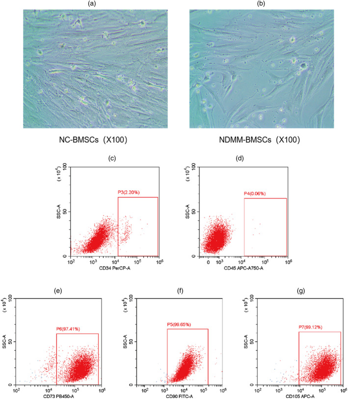
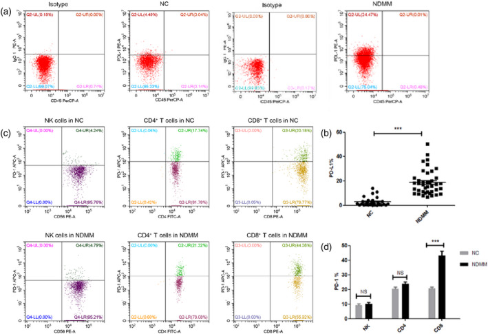
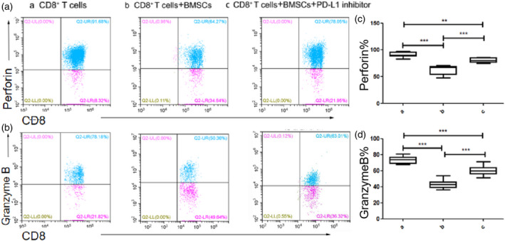
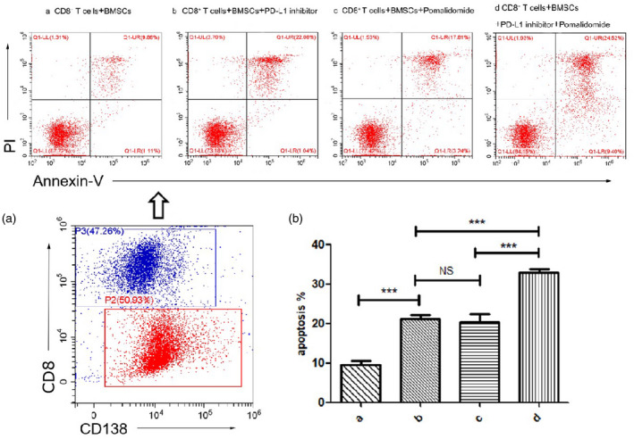
High expression of the inhibitory receptor programmed cell death ligand 1 (PD-L1) on tumor cells and tumor stromal cells have been found to play a key role in tumor immune evasion in several human malignancies. However, the expression of PD-L1 on bone marrow mesenchymal stem cells (BMSCs) and whether the programmed cell death 1 (PD-1)/PD-L1 signal pathway is involved in the BMSCs versus T cell immune response in multiple myeloma (MM) remains poorly defined. In this study, we explored the expression of PD-L1 on BMSCs from newly diagnosed MM (NDMM) patients and the role of PD-1/PD-L1 pathway in BMSC-mediated regulation of CD8+ T cells. The data showed that the expression of PD-L1 on BMSCs in NDMM patients was significantly increased compared to that in normal controls (NC) (18·81 ± 1·61 versus 2·78± 0·70%; P < 0·001). Furthermore, the PD-1 expression on CD8+ T cells with NDMM patients was significantly higher than that in normal controls (43·22 ± 2·98 versus 20·71 ± 1·08%; P < 0·001). However, there was no significant difference in PD-1 expression of CD4+ T cells and natural killer (NK) cells between the NDMM and NC groups. Additionally, the co-culture assays revealed that BMSCs significantly suppressed CD8+ T cell function. However, the PD-L1 inhibitor effectively reversed BMSC-mediated suppression in CD8+ T cells. We also found that the combination of PD-L1 inhibitor and pomalidomide can further enhance the killing effect of CD8+ T cells on MM cells. In summary, our findings demonstrated that BMSCs in patients with MM may induce apoptosis of CD8+ T cells through the PD-1/PD-L1 axis and inhibit the release of perforin and granzyme B from CD8+ T cells to promote the immune escape of MM.
Keywords: CD8+ T cells; PD-1/PD-L1; bone marrow mesenchymal stem cells (BMSCs); multiple myeloma (MM); pomalidomide.
© 2021 British Society for Immunology.
Conflict of interest statement
All authors report no conflicts of interest.
Figures
Similar articles
-
Bone marrow-derived mesenchymal stem cells promote cell proliferation of multiple myeloma through inhibiting T cell immune responses via PD-1/PD-L1 pathway.Cell Cycle. 2018;17(7):858-867. doi: 10.1080/15384101.2018.1442624. Epub 2018 May 21. Cell Cycle. 2018. PMID: 29493401 Free PMC article.
-
Tumor-associated macrophages regulate the function of cytotoxic T lymphocyte through PD-1/PD-L1 pathway in multiple myeloma.Cancer Med. 2022 Dec;11(24):4838-4848. doi: 10.1002/cam4.4814. Epub 2022 May 20. Cancer Med. 2022. PMID: 35593325 Free PMC article.
-
PD-L1/PD-1 Pattern of Expression Within the Bone Marrow Immune Microenvironment in Smoldering Myeloma and Active Multiple Myeloma Patients.Front Immunol. 2021 Jan 8;11:613007. doi: 10.3389/fimmu.2020.613007. eCollection 2020. Front Immunol. 2021. PMID: 33488620 Free PMC article.
-
Activation of NK cells and disruption of PD-L1/PD-1 axis: two different ways for lenalidomide to block myeloma progression.Oncotarget. 2017 Apr 4;8(14):24031-24044. doi: 10.18632/oncotarget.15234. Oncotarget. 2017. PMID: 28199990 Free PMC article. Review.
-
Role of the tumor microenvironment in PD-L1/PD-1-mediated tumor immune escape.Mol Cancer. 2019 Jan 15;18(1):10. doi: 10.1186/s12943-018-0928-4. Mol Cancer. 2019. PMID: 30646912 Free PMC article. Review.
Cited by
-
Potential functions and therapeutic implications of glioma-resident mesenchymal stem cells.Cell Biol Toxicol. 2023 Jun;39(3):853-866. doi: 10.1007/s10565-023-09808-7. Epub 2023 May 3. Cell Biol Toxicol. 2023. PMID: 37138122 Review.
-
Mesenchymal stromal cells in bone marrow niche of patients with multiple myeloma: a double-edged sword.Cancer Cell Int. 2025 Mar 26;25(1):117. doi: 10.1186/s12935-025-03741-x. Cancer Cell Int. 2025. PMID: 40140850 Free PMC article. Review.
-
CXCL12/CXCR4 axis supports mitochondrial trafficking in tumor myeloma microenvironment.Oncogenesis. 2022 Jan 21;11(1):6. doi: 10.1038/s41389-022-00380-z. Oncogenesis. 2022. PMID: 35064098 Free PMC article.
-
Exosomal miR-483-5p in Bone Marrow Mesenchymal Stem Cells Promotes Malignant Progression of Multiple Myeloma by Targeting TIMP2.Front Cell Dev Biol. 2022 Mar 1;10:862524. doi: 10.3389/fcell.2022.862524. eCollection 2022. Front Cell Dev Biol. 2022. PMID: 35300408 Free PMC article.
-
Engagement of Mesenchymal Stromal Cells in the Remodeling of the Bone Marrow Microenvironment in Hematological Cancers.Biomolecules. 2023 Nov 24;13(12):1701. doi: 10.3390/biom13121701. Biomolecules. 2023. PMID: 38136573 Free PMC article. Review.
References
Publication types
MeSH terms
Substances
LinkOut - more resources
Full Text Sources
Other Literature Sources
Medical
Research Materials
