. 2021 Apr 29;184(9):2362-2371.e9.
doi: 10.1016/j.cell.2021.02.042. Epub 2021 Feb 23.

SARS-CoV-2 501Y.V2 variants lack higher infectivity but do have immune escape

Affiliations

SARS-CoV-2 501Y.V2 variants lack higher infectivity but do have immune escape

Qianqian Li et al. Cell. .

Abstract

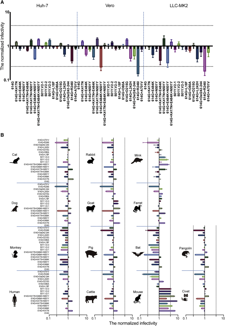
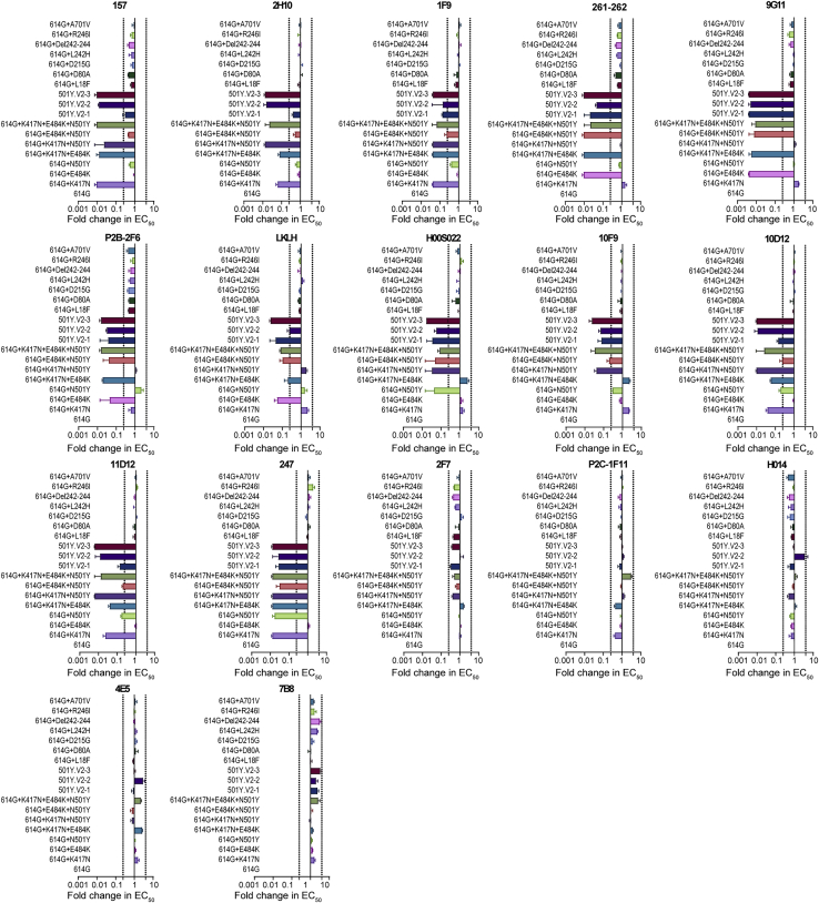
The 501Y.V2 variants of SARS-CoV-2 containing multiple mutations in spike are now dominant in South Africa and are rapidly spreading to other countries. Here, experiments with 18 pseudotyped viruses showed that the 501Y.V2 variants do not confer increased infectivity in multiple cell types except for murine ACE2-overexpressing cells, where a substantial increase in infectivity was observed. Notably, the susceptibility of the 501Y.V2 variants to 12 of 17 neutralizing monoclonal antibodies was substantially diminished, and the neutralization ability of the sera from convalescent patients and immunized mice was also reduced for these variants. The neutralization resistance was mainly caused by E484K and N501Y mutations in the receptor-binding domain of spike. The enhanced infectivity in murine ACE2-overexpressing cells suggests the possibility of spillover of the 501Y.V2 variants to mice. Moreover, the neutralization resistance we detected for the 501Y.V2 variants suggests the potential for compromised efficacy of monoclonal antibodies and vaccines.

Keywords: 501Y.V2; E484K; K417N; N501Y; SARS-CoV-2; immune escape; infectivity; mutation; neutralizing antibody; receptor binding region.


Conflict of interest statement

Declaration of interests The authors declare no competing interests.

Figures

None
Graphical abstract
Figure 1
Figure 1
Illustration of 501Y.V2-related pseudotyped viruses (A) All the 501Y.V2-related mutation sites. (B) 501Y.V2-1. (C) 501Y.V2-2. (D) 501Y.V2-3.
Figure 2
Figure 2
Infectivity analysis of mutant pseudotyped viruses (A) Infection assays with the 18 501Y.V2-related mutant pseudotyped viruses with the three indicated cell lines, all of which are known to be susceptible to SARS-CoV-2. (B) Infection assays for a set of 14 HEK293T cell lines each expressing the indicated mammalian ortholog of ACE2. The infectivity of the reference 614G variant was used as a control (i.e., the infectivity of other 17 variants in each experiment was normalized to values detected for the reference 614G variant). Data are the means ± SEM of six independent experiments. The dashed lines indicate the threshold value of a 4-fold difference in infectivity. See also Figure S1.
Figure S1
Figure S1
The expression levels of the various mammalian ACE2 orthologs on the surface of transfected HEK293T cells, related to Figure 2 The cell surface expression of the FLAG-tagged ACE2 orthologs was assessed by flow cytometry. The PE-A+ value in the upright corner represents the percentage of ACE2-expressing cells.
Figure 3
Figure 3
Analysis of antigenicity of 501Y.V2 variants using a panel of neutralizing monoclonal antibodies Heatmap representation of neutralization reactions using 17 neutralizing monoclonal antibodies—all known to target epitopes in the RBD—against 18 501Y.V2-related mutant pseudotyped viruses; the ratio of EC50 value (for each of the tested antibodies) detected for the reference 614G variant to the EC50 value for each of 501Y.V2-related mutant pseudotyped viruses. Blue and pink represent decreased and increased viral sensitivity to monoclonal antibody neutralization, respectively. Data represent the means of three independent experiments. See also Figures S2 and S3.
Figure S2
Figure S2
Reactivity of pseudotyped viruses with 501Y.V2 related mutations to 17 neutralized monoclonal antibodies, related to Figure 3 The data represent the ratio of the EC50 value for the reference 614G pseudotyped virus to the pseudotyped viruses harboring 501Y.V2 related mutations. Data represent the the means of three independent experiments. The dashed line indicates the threshold value of a 4-fold difference in EC50.
Figure S3
Figure S3
Neutralization curves of the 17 neutralized monoclonal antibodies against the pseudotyped viruses with 501Y.V2 related mutations, related to Figure 3
Figure 4
Figure 4
Analysis of antigenicity of 501Y.V2 variants using a panel of polyclonal antibodies (A) The reactivity of pseudotyped viruses with 501Y.V2-related mutations was assayed against sera from convalescent sera with high-titer polyclonal neutralizing antibodies (“CSC”) from SARS-CoV-2 infection patients and against 3 pooled sera samples (from 9 mice were immunized with RBD [“RBD”]). The data (means of three independent experiments) presented in the heatmap show the ratio of the ED50 value detected for each of the 501Y.V2-related mutant pseudotyped viruses to the value detected for the reference 614G virus. Blue and pink represent decreased and increased viral sensitivity to neutralization by sera, respectively. (B) Summary and inferential statistical analysis of the results for the pseudotyped viruses with 501Y.V2-related mutations. The dashed line represents the mean serum response of the 614G virus. One-way ANOVA and Holm-Sidak’s multiple comparison tests were used to analyze the differences between groups. A p value of <0.05 was considered to be significant. p < 0.05, ∗∗p < 0.01, ∗∗∗p < 0.001.
Figure 5
Figure 5
Testing of longitudinal convalescent sera samples obtained from SARS-CoV-2 infected patients at 2, 5, and 8 months post-onset (A) Heatmap analysis of the ratios of ED50 values of pseudotyped viruses with 501Y.V2-related mutations to the reference 614G virus. Blue and pink represent decreased and increased viral sensitivity to neutralization by sera, respectively. Data represent the means of two independent experiments. (B) Summary and inferential statistical analysis of the results of different mutants. The dashed line represents the mean serum response of the reference 614G virus. One-way ANOVA and Holm-Sidak’s multiple comparisons test were used to analyze the differences between groups. A p-value of <0.05 was considered to be significant. p < 0.05, ∗∗p < 0.01. (C) Analysis of the results of reactions between 501Y.V2-related mutant pseudotyped viruses with longitudinal sera from SARS-CoV-2 infection patients at 2, 5, and 8 months post onset. (D) Model of the S protein trimer (PDB: 6VXX) with human ACE2 and neutralizing antibodies (PDB: 7BZ5, 6XEY). K417 forms hydrogen bonds with the main chain of N370 in the neighboring S protomer in the closed conformation of the S protein.

Comment in

References

    1. Barnes C.O., West A.P., Jr., Huey-Tubman K.E., Hoffmann M.A.G., Sharaf N.G., Hoffman P.R., Koranda N., Gristick H.B., Gaebler C., Muecksch F. Structures of Human Antibodies Bound to SARS-CoV-2 Spike Reveal Common Epitopes and Recurrent Features of Antibodies. Cell. 2020;182:828–842.e16. - PMC - PubMed
    1. Baum A., Ajithdoss D., Copin R., Zhou A., Lanza K., Negron N., Ni M., Wei Y., Mohammadi K., Musser B. REGN-COV2 antibodies prevent and treat SARS-CoV-2 infection in rhesus macaques and hamsters. Science. 2020;370:1110–1115. - PMC - PubMed
    1. Brouwer P.J.M., Caniels T.G., van der Straten K., Snitselaar J.L., Aldon Y., Bangaru S., Torres J.L., Okba N.M.A., Claireaux M., Kerster G. Potent neutralizing antibodies from COVID-19 patients define multiple targets of vulnerability. Science. 2020;369:643–650. - PMC - PubMed
    1. Cao Y., Su B., Guo X., Sun W., Deng Y., Bao L., Zhu Q., Zhang X., Zheng Y., Geng C. Potent Neutralizing Antibodies against SARS-CoV-2 Identified by High-Throughput Single-Cell Sequencing of Convalescent Patients’ B Cells. Cell. 2020;182:73–84.e16. - PMC - PubMed
    1. Crawford K.H.D., Eguia R., Dingens A.S., Loes A.N., Malone K.D., Wolf C.R., Chu H.Y., Tortorici M.A., Veesler D., Murphy M. Protocol and Reagents for Pseudotyping Lentiviral Particles with SARS-CoV-2 Spike Protein for Neutralization Assays. Viruses. 2020;12:5132. - PMC - PubMed

Publication types