

Structure and gating mechanism of the α7 nicotinic acetylcholine receptor
- PMID: 33735609
- PMCID: PMC8135066
- DOI: 10.1016/j.cell.2021.02.049
Structure and gating mechanism of the α7 nicotinic acetylcholine receptor
Abstract
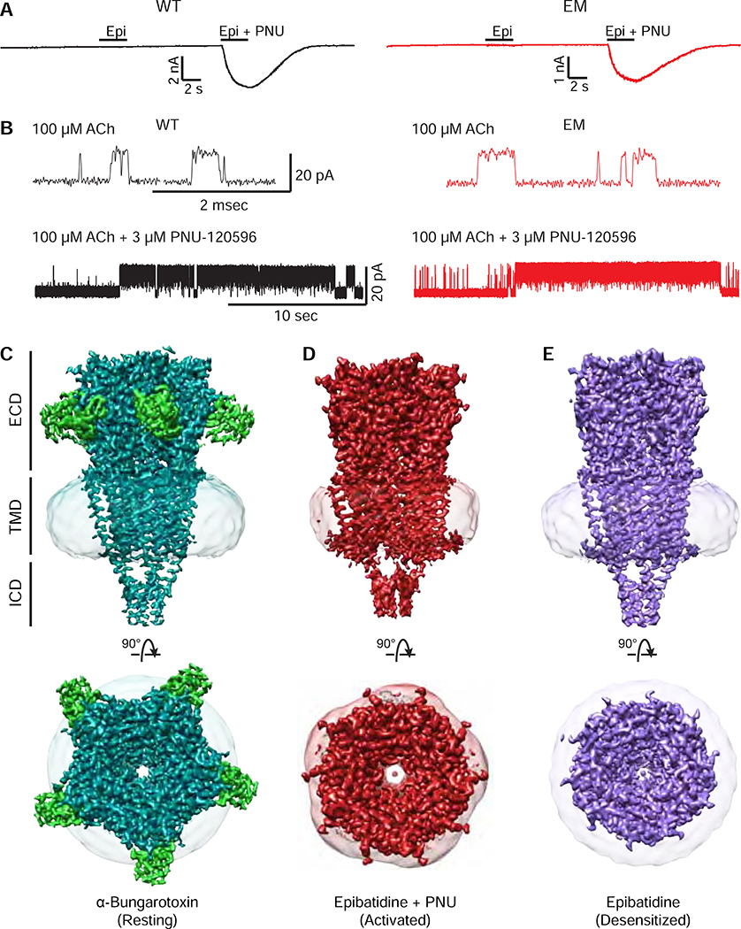
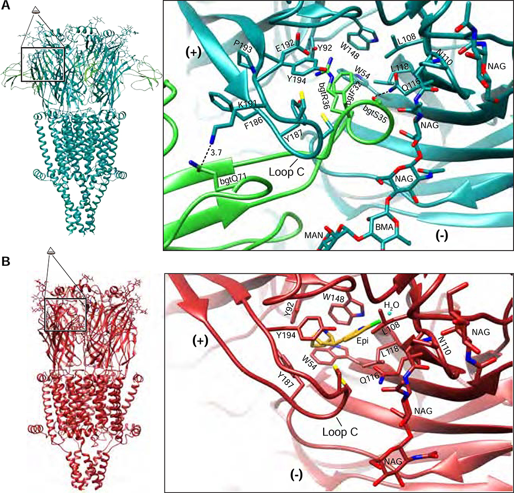
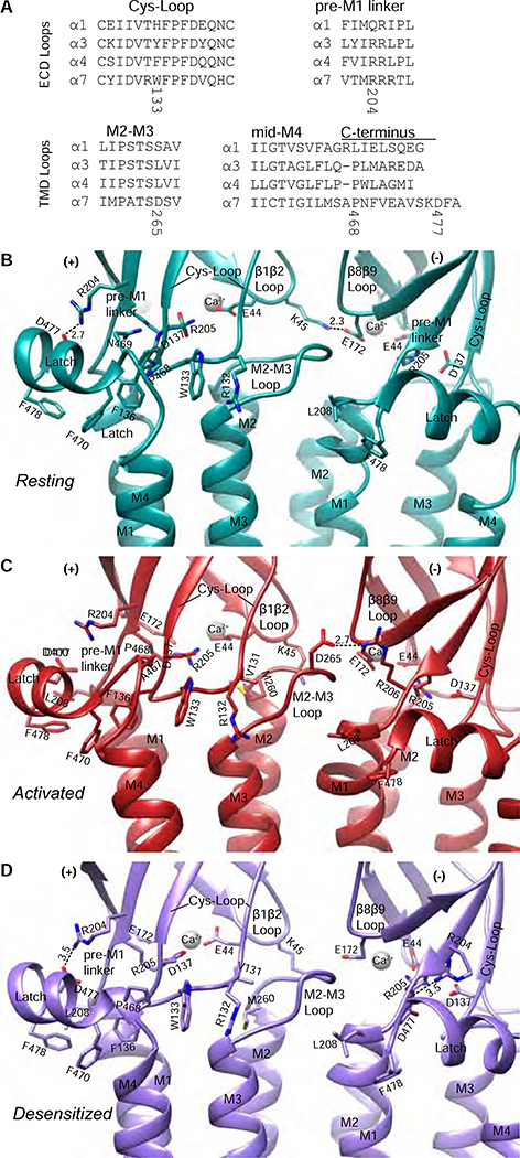
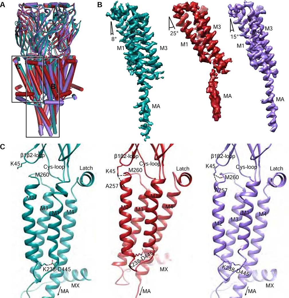
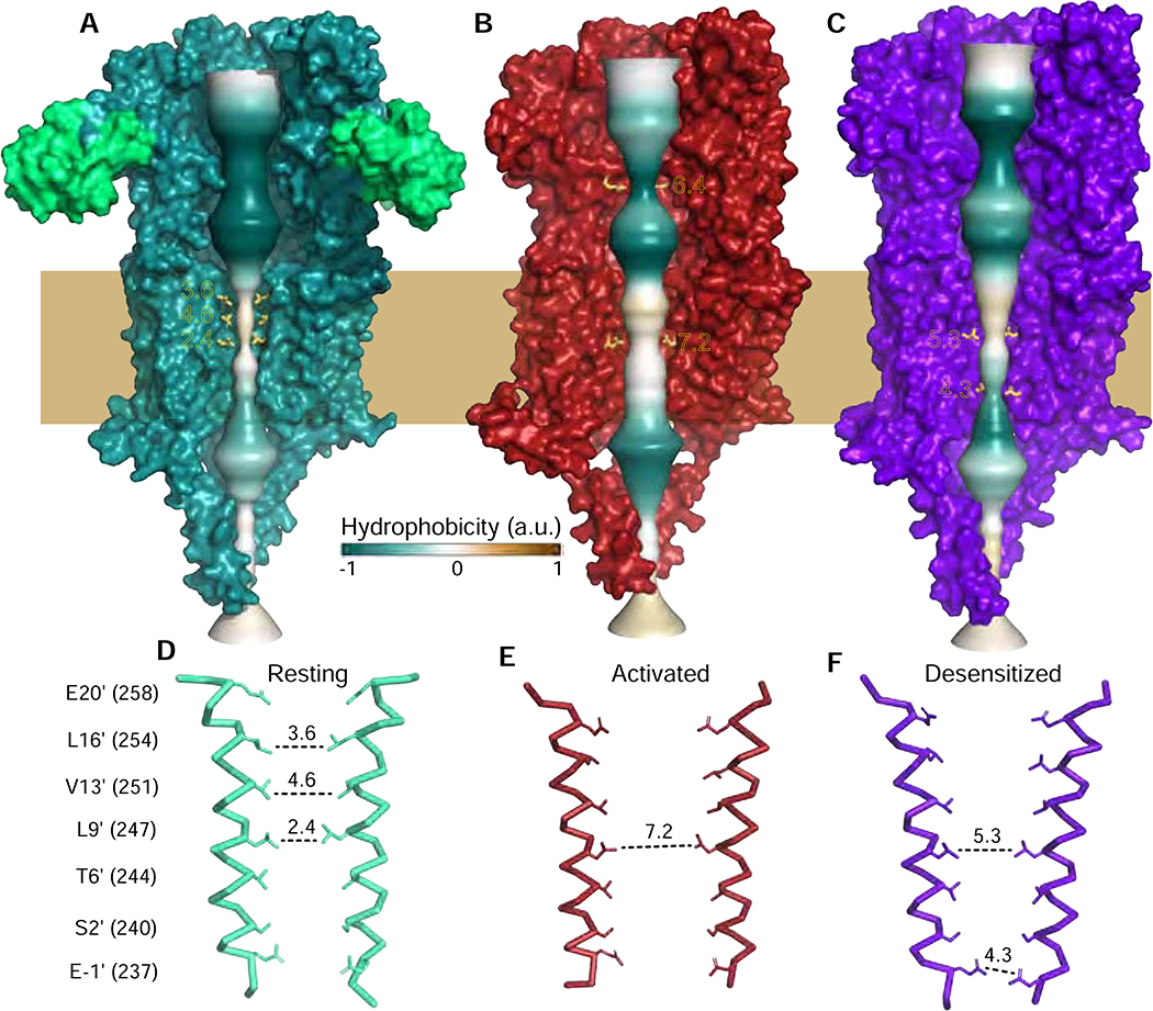
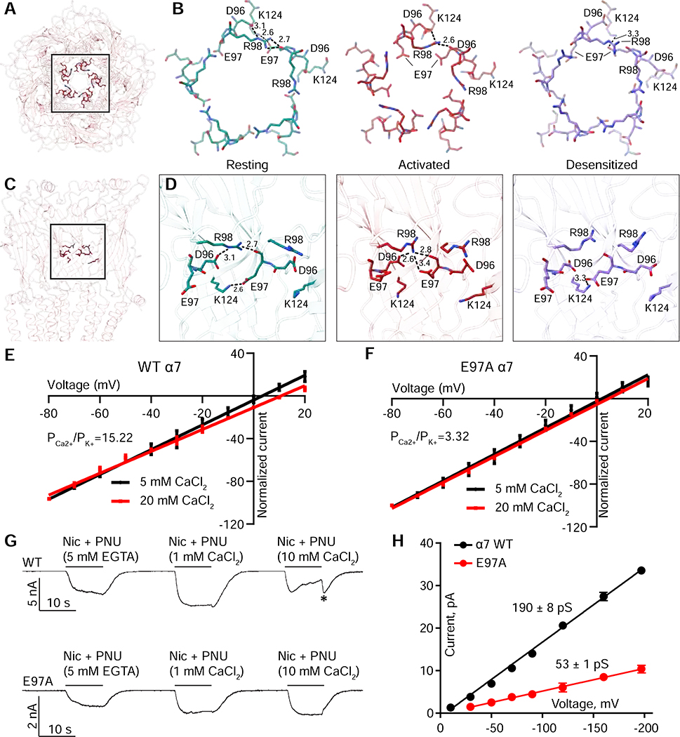
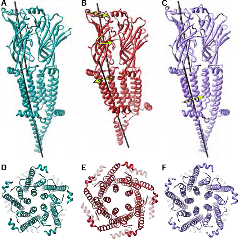
The α7 nicotinic acetylcholine receptor plays critical roles in the central nervous system and in the cholinergic inflammatory pathway. This ligand-gated ion channel assembles as a homopentamer, is exceptionally permeable to Ca2+, and desensitizes faster than any other Cys-loop receptor. The α7 receptor has served as a prototype for the Cys-loop superfamily yet has proven refractory to structural analysis. We present cryo-EM structures of the human α7 nicotinic receptor in a lipidic environment in resting, activated, and desensitized states, illuminating the principal steps in the gating cycle. The structures also reveal elements that contribute to its function, including a C-terminal latch that is permissive for channel opening, and an anionic ring in the extracellular vestibule that contributes to its high conductance and calcium permeability. Comparisons among the α7 structures provide a foundation for mapping the gating cycle and reveal divergence in gating mechanisms in the Cys-loop receptor superfamily.
Keywords: Cys-loop receptor; acetylcholine receptor; cryo-EM; ion channel; ligand-gated ion channel; nicotinic receptor; α7.
Copyright © 2021 Elsevier Inc. All rights reserved.
Conflict of interest statement
Declaration of interests The authors declare no competing interests.
Figures
Similar articles
-
A human-specific, truncated α7 nicotinic receptor subunit assembles with full-length α7 and forms functional receptors with different stoichiometries.J Biol Chem. 2018 Jul 6;293(27):10707-10717. doi: 10.1074/jbc.RA117.001698. Epub 2018 May 21. J Biol Chem. 2018. PMID: 29784875 Free PMC article.
-
The coupling interface and pore domain codetermine the single-channel activity of the α7 nicotinic receptor.Neuropharmacology. 2015 Aug;95:448-58. doi: 10.1016/j.neuropharm.2015.04.010. Epub 2015 Apr 20. Neuropharmacology. 2015. PMID: 25908400
-
An allosteric binding site of the α7 nicotinic acetylcholine receptor revealed in a humanized acetylcholine-binding protein.J Biol Chem. 2018 Feb 16;293(7):2534-2545. doi: 10.1074/jbc.M117.815316. Epub 2017 Dec 13. J Biol Chem. 2018. PMID: 29237730 Free PMC article.
-
In Silico Modeling of the α7 Nicotinic Acetylcholine Receptor: New Pharmacological Challenges Associated with Multiple Modes of Signaling.Mini Rev Med Chem. 2020;20(10):841-864. doi: 10.2174/1389557520666200130105256. Mini Rev Med Chem. 2020. PMID: 32000651 Free PMC article. Review.
-
Combining Mutations and Electrophysiology to Map Anesthetic Sites on Ligand-Gated Ion Channels.Methods Enzymol. 2018;602:369-389. doi: 10.1016/bs.mie.2018.01.014. Epub 2018 Feb 28. Methods Enzymol. 2018. PMID: 29588039 Free PMC article. Review.
Cited by
-
Analogs of α-conotoxin PnIC selectively inhibit α7β2- over α7-only subtype nicotinic acetylcholine receptors via a novel allosteric mechanism.FASEB J. 2024 Jan;38(1):e23374. doi: 10.1096/fj.202302079. FASEB J. 2024. PMID: 38161283 Free PMC article.
-
Pursuing High-Resolution Structures of Nicotinic Acetylcholine Receptors: Lessons Learned from Five Decades.Molecules. 2021 Sep 23;26(19):5753. doi: 10.3390/molecules26195753. Molecules. 2021. PMID: 34641297 Free PMC article. Review.
-
Structural mechanisms of α7 nicotinic receptor allosteric modulation and activation.Cell. 2024 Feb 29;187(5):1160-1176.e21. doi: 10.1016/j.cell.2024.01.032. Epub 2024 Feb 20. Cell. 2024. PMID: 38382524 Free PMC article.
-
Differential Activation and Desensitization States Promoted by Noncanonical α7 Nicotinic Acetylcholine Receptor Agonists.J Pharmacol Exp Ther. 2022 Nov;383(2):157-171. doi: 10.1124/jpet.122.001354. Epub 2022 Sep 2. J Pharmacol Exp Ther. 2022. PMID: 36279397 Free PMC article.
-
Structural basis for allosteric agonism of human α7 nicotinic acetylcholine receptors.Cell Discov. 2025 Apr 8;11(1):35. doi: 10.1038/s41421-025-00788-y. Cell Discov. 2025. PMID: 40195322 Free PMC article.
References
-
- Aldea M, Mulet J, Sala S, Sala F, and Criado M (2007). Non-charged amino acids from three different domains contribute to link agonist binding to channel gating in alpha7 nicotinic acetylcholine receptors. J Neurochem 103, 725–735. - PubMed
-
- Alves DS, Castello-Banyuls J, Faura CC, and Ballesta JJ (2011). An extracellular RRR motif flanking the M1 transmembrane domain governs the biogenesis of homomeric neuronal nicotinic acetylcholine receptors. FEBS Lett 585, 1169–1174. - PubMed
Publication types
MeSH terms
Substances
Grants and funding
LinkOut - more resources
Full Text Sources
Other Literature Sources
Miscellaneous
