Review
. 2021 Aug:63:78-85.
doi: 10.1016/j.cbpa.2021.01.014. Epub 2021 Mar 15.

Imaging-guided nanomedicine development

Affiliations
Review

Imaging-guided nanomedicine development

Aurora Bernal et al. Curr Opin Chem Biol. 2021 Aug.

Abstract

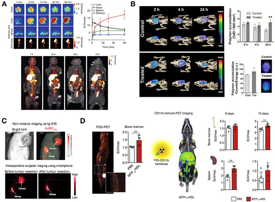
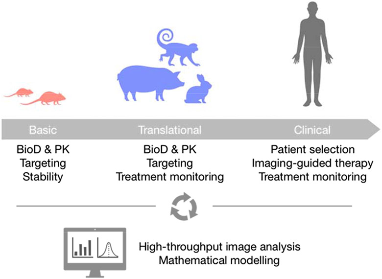
Nanomedicine research is an active field that produces thousands of studies every year. However, translation of nanotherapeutics to the clinic has yet to catch up with such a vast output. In recent years, the need to better understand nanomedicines' in vivo behavior has been identified as one of the major challenges for efficient clinical translation. In this context, noninvasive imaging offers attractive solutions to provide valuable information about nanomedicine biodistribution, pharmacokinetics, stability, or therapeutic efficacy. Here, we review the latest imaging approaches used in the development of therapeutic nanomedicines, discuss why these strategies bring added value along the translational pipeline, and give a perspective on future advances in the field.


Conflict of interest statement

Declaration of competing interest The authors declare the following financial interests/personal relationships which may be considered as potential competing interests: WJMM is founder of Trained Therapeutix Discovery.

Figures

Figure 1.
Figure 1.. Non-invasive imaging applications in nanomedicine development.
A) Biodistribution and pharmacokinetics. The tissue distribution and clearance kinetics of a TRAF6 inhibitor-loaded nanobiologic was evaluated in non-human primates by PET/MRI. The formulation was radiolabeled with 89Zr and monitored dynamically for the first hour after administration (top left). Quantitative data could be derived from selected tissues (top right). Subsequently, static scans were performed at 24, 48 and 72 hours post injection (bottom). Adapted from Lameijer et al. [4]. B) Targeting efficiency. A sonoporation treatment to enhance delivery across the BBB was evaluated by in vivo FMT/CT imaging using fluorophore-labeled nanoparticles. A significant increase in the florescent signal was measured 24 hours post-administration in treated animals compared to controls (top right). The results were corroborated ex vivo by fluorescence reflectance imaging of explanted brains (bottom right). Adapted from May et al. [19]. C) Imaging-guided therapy. An ultra-small gold quantum cluster nanoparticle (AuQC705), detectable by near-infrared fluorescence, CT and MRI, was successfully employed to guide tumor resection by fluorescence imaging using a portable smartphone imaging system prototype. Adapted from Yang et al. [39]. D) Treatment monitoring. Non-invasive imaging can be used to monitor nanomedicine treatment efficacy and its underlying mechanisms of action. A trained-immunity promoting nanobiologic (MTP10-HDL) was developed as a novel anticancer therapy. Its effects on immune response activation were monitored by PET imaging of metabolic activation in the bone marrow using 18F-FDG (FDG-PET, left) and myelopoiesis in bone marrow and spleen using a radiolabeled nanobody (CD11b immuno-PET, right). Adapted from Priem at al. [17].
Figure 2.
Figure 2.. Integration of imaging along the translational pipeline.
Non-invasive imaging can be integrated at different stages of a nanoformulation’s development. At the early stages, imaging-based screening of promising candidates can be performed to elucidate their in vivo behavior in terms of biodistribution (BioD), pharmacokinetics (PK), targeting or stability. In addition to assessing in vivo behavior, translational studies in large animal models can benefit from the integration of non-invasive imaging to longitudinally investigate treatment response using limited group sizes. Finally, in the clinic, imaging-based protocols can aid in selecting amenable patients, guiding therapy and monitoring response. At all stages, AI-based image analyses will be of paramount importance to generate quality data and facilitate mathematical modelling in order to understand and possibly predict nanomedicines’ performance.

References

    1. Germain M, Caputo F, Metcalfe S, Tosi G, Spring K, Åslund AKO, Pottier A, Schiffelers R, Ceccaldi A, Schmid R: Delivering the power of nanomedicine to patients today. J Control Release 2020, 326:164–171. - PMC - PubMed
    1. Anselmo AC, Mitragotri S: Nanoparticles in the clinic: An update. Bioeng Transl Med 2019, 4:e10143. - PMC - PubMed
    1. Binderup T, Duivenvoorden R, Fay F, van Leent MMT, Malkus J, Baxter S, Ishino S, Zhao Y, Sanchez-Gaytan B, Teunissen AJP, et al.: Imaging-assisted nanoimmunotherapy for atherosclerosis in multiple species. Sci Transl Med 2019, 11:eaaw7736.

      (••) In this study, we implemented an imaging-based approach to translate a nanoimmunotherapy from small to large animals that included evaluation of in vivo behavior by PET/CT and PET/MRI, as well as treatment monitoring by PET/MRI.

    1. Lameijer M, Binderup T, Van Leent MMT, Senders ML, Fay F, Malkus J, Sanchez-Gaytan BL, Teunissen AJP, Karakatsanis N, Robson P, et al.: Efficacy and safety assessment of a TRAF6-targeted nanoimmunotherapy in atherosclerotic mice and non-human primates. Nat Biomed Eng 2018, 2:279–292. - PMC - PubMed
    1. Braza MS, van Leent MMT, Lameijer M, Sanchez-Gaytan BL, Arts RJW, Pérez-Medina C, Conde P, Garcia MR, Gonzalez-Perez M, Brahmachary M, et al.: Inhibiting Inflammation with Myeloid Cell-Specific Nanobiologics Promotes Organ Transplant Acceptance. Immunity 2018, 49:819–828.e6. - PMC - PubMed

Publication types

LinkOut - more resources