

Cerebrovascular reactivity in retinal vasculopathy with cerebral leukoencephalopathy and systemic manifestations
- PMID: 33736510
- PMCID: PMC7983338
- DOI: 10.1177/0271678X20929430
Cerebrovascular reactivity in retinal vasculopathy with cerebral leukoencephalopathy and systemic manifestations
Abstract
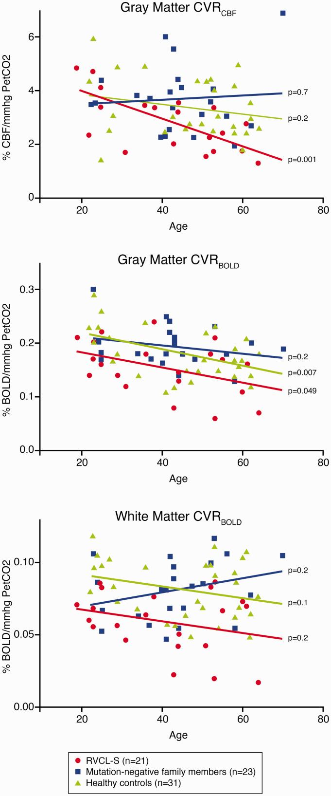
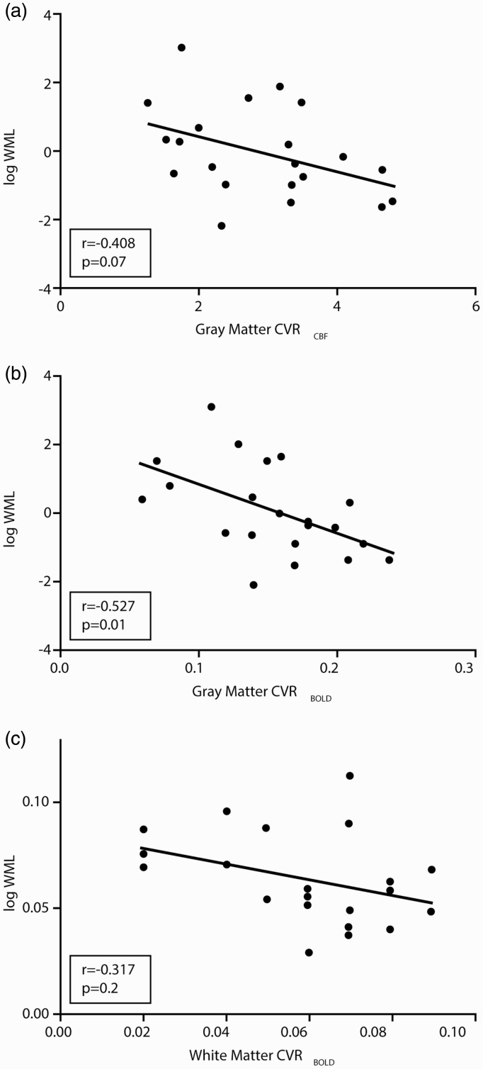
Retinal Vasculopathy with Cerebral Leukoencephalopathy and Systemic manifestations (RVCL-S) is a small vessel disease caused by TREX1 mutations. RVCL-S is characterized by retinal vasculopathy and brain white matter lesions with and without contrast enhancement. We aimed to investigate cerebrovascular reactivity (CVR) in RVCL-S. In this cross-sectional observational study, 21 RVCL-S patients, 23 mutation-negative family members, and 31 healthy unrelated controls were included. CVR to a hypercapnic challenge was measured using dual-echo arterial spin labeling magnetic resonance imaging. Stratified analyses based on age were performed. We found that CVR was decreased in gray and white matter of RVCL-S patients compared with family members and healthy controls (ANCOVA; P < 0.05 for all comparisons). This was most noticeable in RVCL-S patients aged ≥40 years (ANCOVA, P < 0.05 for all comparisons). In RVCL-S patients aged < 40 years, only CVR in white matter was lower when compared to healthy controls (P < 0.05). Gray matter CVR was associated with white matter lesion volume in RVCL-S patients (r = -0.527, P = 0.01). In conclusion, impaired cerebrovascular reactivity may play an important role in the pathophysiology of RVCL-S and may be an useful early biomarker of cerebrovascular disease severity.
Keywords: Cerebrovascular circulation; cerebrovascular disorders; leukoencephalopathies; magnetic resonance imaging; mutation.
Conflict of interest statement
Figures
References
-
- Richards A, Van den Maagdenberg AM, Jen JC, et al.. C-terminal truncations in human 3'-5' DNA exonuclease TREX1 cause autosomal dominant retinal vasculopathy with cerebral leukodystrophy. Nat Genet 2007; 39: 1068–1070. - PubMed
-
- Jen J, Cohen AH, Yue Q, et al.. Hereditary endotheliopathy with retinopathy, nephropathy, and stroke (HERNS). Neurology 1997; 49: 1322–1330. - PubMed
-
- Terwindt GM, Haan J, Ophoff RA, et al.. Clinical and genetic analysis of a large Dutch family with autosomal dominant vascular retinopathy, migraine and Raynaud’s phenomenon. Brain 1998; 121: 303–316. - PubMed
Publication types
MeSH terms
Substances
LinkOut - more resources
Full Text Sources
