. 2021 Mar 18;18(1):13.
doi: 10.1186/s12987-021-00247-2.

Astrocyte-specific hypoxia-inducible factor 1 (HIF-1) does not disrupt the endothelial barrier during hypoxia in vitro

Affiliations

Astrocyte-specific hypoxia-inducible factor 1 (HIF-1) does not disrupt the endothelial barrier during hypoxia in vitro

Julia Baumann et al. Fluids Barriers CNS. .

Abstract

Background: Astrocytes (AC) are essential for brain homeostasis. Much data suggests that AC support and protect the vascular endothelium, but increasing evidence indicates that during injury conditions they may lose their supportive role resulting in endothelial cell activation and BBB disturbance. Understanding the triggers that flip this switch would provide invaluable information for designing new targets to modulate the brain vascular compartment. Hypoxia-inducible factor-1 (HIF-1) has long been assumed to be a culprit for barrier dysfunction as a number of its target genes are potent angiogenic factors. Indeed AC themselves, reservoirs of an array of different growth factors and molecules, are frequently assumed to be the source of such molecules although direct supporting evidence is yet to be published. Being well known reservoirs of HIF-1 dependent angiogenic molecules, we asked if AC HIF-1 dependent paracrine signaling drives brain EC disturbance during hypoxia.

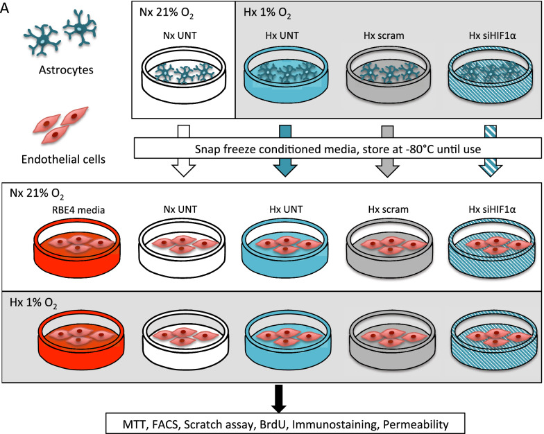
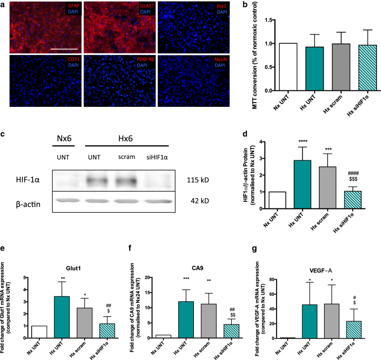
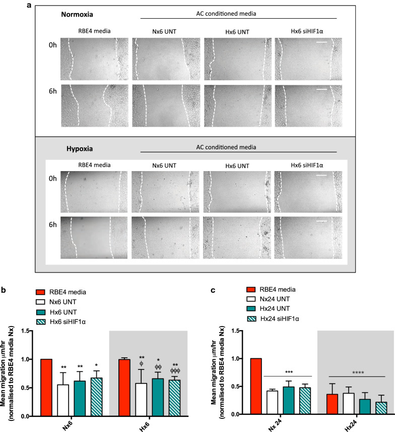
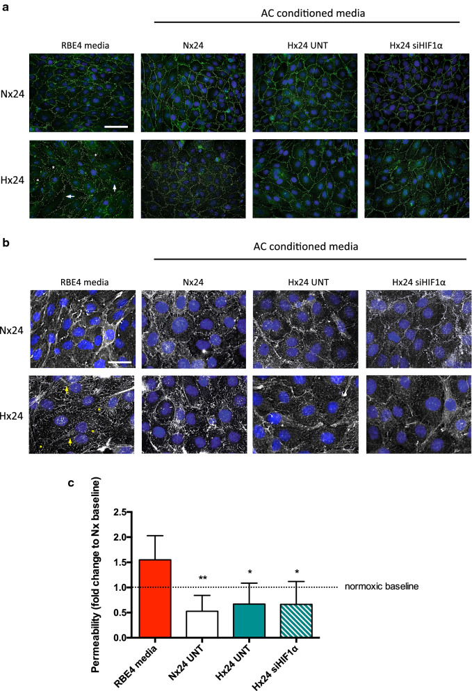
Methods: First we collected conditioned media from control and siRNA-mediated HIF-1 knockdown primary rat AC that had been exposed to normoxic or hypoxic conditions. The conditioned media was then used to culture normoxic and hypoxic (1% O2) rat brain microvascular EC (RBE4) for 6 and 24 h. Various activation parameters including migration, proliferation and cell cycling were assessed and compared to untreated controls. In addition, tight junction localization and barrier stability per se (via permeability assay) was evaluated.

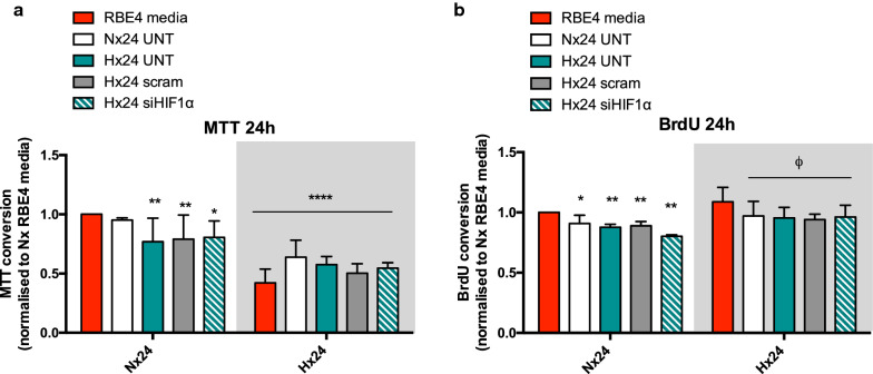
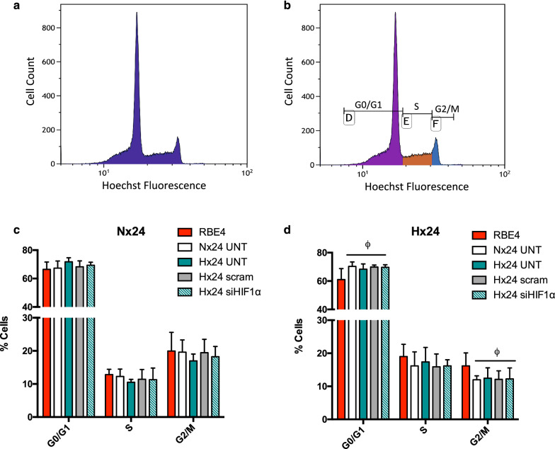
Results: AC conditioned media maintained both normoxic and hypoxic EC in a quiescent state by suppressing EC metabolic activity and proliferation. By FACs we observed reduced cell cycling with an increased number of cells in G0 phase and reduced cell numbers in M phase compared to controls. EC migration was also blocked by AC conditioned media and in correlation hypoxic tight junction organization and barrier functionality was improved. Surprisingly however, AC HIF-1 deletion did not impact EC responses or barrier stability during hypoxia.

Conclusions: This study demonstrates that AC HIF-1 dependent paracrine signaling does not contribute to AC modulation of EC barrier function under normoxic or hypoxic conditions. Thus other cell types likely mediate EC permeability in stress scenarios. Our data does however highlight the continuous protective effect of AC on the barrier endothelium. Exploring these protective mechanisms in more detail will provide essential insight into ways to prevent barrier disturbance during injury and disease.

Keywords: Astrocytes; Barrier stability; Endothelial activation; HIF-1; In vitro BBB; Primary culture.


Conflict of interest statement

The authors declare that they have no competing interests.

Figures

Fig. 1
Fig. 1
Schematic of AC-CM experiments. Graphical overview of experimental setup using primary AC (dark green) and the RBE4 EC cell line (red). Media was collected from normoxic (white) or hypoxic untreated (UNT, teal), scrambled transfected (scram, grey) or siHIF-1α transfected AC (si-HIF1α, teal-dashed) and snap frozen before use. Thawed AC-CM was transferred to confluent RBE4 immediately before exposure. All experiments were compared to RBE4 grown in their own culture media (RBE4 media, red)
Fig. 2
Fig. 2
Confirmation of HIF-1α deletion in primary astrocytes. a Micrographs showing primary rat astrocytes immunostained for GLAST, GFAP, Iba1, CD31, PDGFRβ and NeuN expression, counterstained with DAPI (blue). Scale bar = 200 nm. b MTT conversion of primary AC after 24 h normoxic or hypoxic exposure with HIF-1 KD (siHIF1α) or untreated (UNT) and scrambled controls (scram). c Representative Western blot and d quantification of HIF-1α KD efficiency in primary ACs after 6 h hypoxia. Messenger RNA levels of HIF-1 target genes Glut1, CA9 and VEGF-A in primary KD ACs compared to normoxic and hypoxic controls after 24 h hypoxia (eg). Students t-test and 2way ANOVA, mean ± SD, n = 4–6, *p < 0.05, **p < 0.01, ***p < 0.001, ****p < 0.0001 compared to Nx UNT, #p < 0.05, ##p < 0.01 ####p < 0.0001 to Hx UNT, $p < 0.05, $$$p < 0.001 compared to Hx scram
Fig. 3
Fig. 3
Astrocyte HIF-1 paracrine signalling does not affect endothelial cell metabolic activity and proliferation. Graphical representation of EC a mitochondrial activity and b proliferation as measured by BrdU incorporation after 24 h exposure. EC were exposed to normoxic or hypoxic conditions with AC-CM or their own RBE4 media. Data is presented in comparison to baseline controls (normoxic RBE4 media control; red bar). 2way ANOVA, mean ± SD, n = 4–6, *p < 0.05, **p < 0.01, ****p < 0.0001 compared to RBE4 media Nx, ϕp < 0.05 to RBE4 media Hx
Fig. 4
Fig. 4
AC conditioned media suppresses hypoxia-induced EC cell cycling. a Representative histogram of FACS performed on EC labeled with the DNA dye Hoechst. b Example of single cell selective gating during cell cycle phases G0/G1, S and G2/M. Quantification and graphical representation of cell percentages in the individual cell cycle phases during 24 h c normoxic and d hypoxic exposures with AC-CM. Students t-test, mean ± SD, n = 3–5, ϕp < 0.05 compared to RBE4 media Hx
Fig. 5
Fig. 5
AC HIF-1 KD does not alter EC migration. a Representative images of scratched confluent EC monolayers at 0 h and after 6 h exposure. Scale bar = 100 μm. EC were scratched and subsequently treated with RBE4 media or normoxic and hypoxic AC-CM, then exposed to normoxia or hypoxia for 6 h or 24 h. After quantification mean migration is graphed as µm/h for b 6 h and c 24 h. 2way ANOVA, mean ± SD, n = 3, *p < 0.05, **p < 0.01 ***p < 0.001, ****p < 0.0001 compared to RBE4 media Nx, ϕp < 0.05, ϕϕp < 0.01, ϕϕϕp < 0.001 to RBE4 media Hx
Fig. 6
Fig. 6
Protection of barrier functionality by AC-CM is independent of HIF-1. a Immunostaining of confluent RBE4 labeled for ZO-1 (green) and cell nuclei (DAPI, blue). Cells were cultured in RBE4 or AC conditioned media under normoxia or hypoxia for 24 h. Hypoxia-induced disruption of ZO-1 (arrows) and inter-endothelial gap formation (asterisks) is seen at cell–cell borders. Scale bar = 50 μm. b Phalloidin (white) and DAPI stain (blue) of confluent RBE4 monolayers after 24 h normoxic or hypoxic exposure with AC-CM or RBE4 media. Arrows indicate hypoxic stress fiber formation, asterisks highlight inter-endothelial gap formation. Enhanced images, scale bar = 25 μm. c Permeability assays were performed on confluent RBE4 on Transwell inserts. Cells were treated with RBE4 media or AC-CM for 24 h. Results are compared to normoxic baseline (dotted line). 2way ANOVA, mean ± SD, n = 3–4, *p < 0.05, **p < 0.01 compared to RBE4 media

Similar articles

Cited by

References

    1. Sofroniew MV, Vinters HV. Astrocytes: biology and pathology. Acta Neuropathol. 2010;119(1):7–35. - PMC - PubMed
    1. Hertz L, Chen Y. Importance of astrocytes for potassium ion (K(+)) homeostasis in brain and glial effects of K(+) and its transporters on learning. Neurosci Biobehav Rev. 2016;71:484–505. - PubMed
    1. Ioannou MS, Jackson J, Sheu SH, Chang CL, Weigel AV, Liu H, et al. Neuron-astrocyte metabolic coupling protects against activity-induced fatty acid toxicity. Cell. 2019;177(6):1522.e14–1535.e14. - PubMed
    1. Huang SF, Fischer S, Koshkin A, Laczko E, Fischer D, Ogunshola OO. Cell-specific metabolomic responses to injury: novel insights into blood–brain barrier modulation. Sci Rep. 2020;10(1):7760. - PMC - PubMed
    1. McCaffrey G, Staatz WD, Quigley CA, Nametz N, Seelbach MJ, Campos CR, et al. Tight junctions contain oligomeric protein assembly critical for maintaining blood–brain barrier integrity in vivo. J Neurochem. 2007;103(6):2540–2555. - PubMed

Substances

LinkOut - more resources