

Real-time ultrasound for tip location of umbilical venous catheter in neonates: a pre/post intervention study
- PMID: 33736669
- PMCID: PMC7977571
- DOI: 10.1186/s13052-021-01014-7
Real-time ultrasound for tip location of umbilical venous catheter in neonates: a pre/post intervention study
Abstract
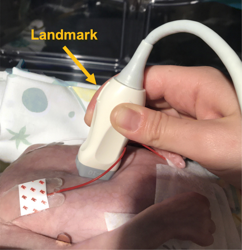
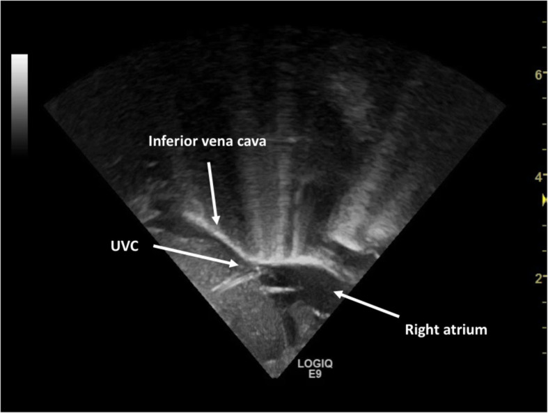
Background: Recent guidelines advocate the use of real-time ultrasound to locate umbilical venous catheter tip. So far, training programs are not well established.
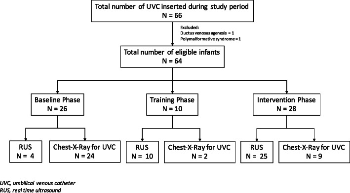
Methods: A pre/post interventional study was carried out in our tertiary neonatal intensive care unit centre to evaluate the efficacy of a training protocol in the use of real-time ultrasound. Primary outcome was the percentage in the use of real-time ultrasound.
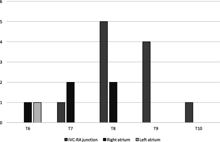
Results: Fifty-four patients were enrolled. The use of real-time ultrasound for tip location significantly increased after the training program (15.3% vs 89.2%, p < 0.0001). After the training the tip of the catheters was more frequently placed at the junction of the inferior vena cava and right atrium (75% vs 30.7%, p = 0.0023). Twenty-two catheters were also evaluated with serial scans during the intervention phase to assess migration rate which was 50%.
Conclusion: a multimodal, targeted training on the use of real-time ultrasound for umbilical venous catheter placement is feasible. Real-time ultrasound is easily teachable, increases the number of umbilical venous catheters placed in a correct position, reduces the number of line manipulations and the need of chest-x-rays.
Keywords: Neonates; Real-time ultrasound; Training; Umbilical venous catheter.
Conflict of interest statement
The authors declare that they have no competing interests.
Figures
References
-
- Shukla H, Ferrara AX. Rapid estimation of insertional length of umbilical catheters in newborns. Am J Dis Child. 1966;140:786–788. - PubMed
MeSH terms
LinkOut - more resources
Full Text Sources
Other Literature Sources
