

Development and Validation of a Deep Learning-Based Model to Distinguish Glioblastoma from Solitary Brain Metastasis Using Conventional MR Images
- PMID: 33737268
- PMCID: PMC8115383
- DOI: 10.3174/ajnr.A7003
Development and Validation of a Deep Learning-Based Model to Distinguish Glioblastoma from Solitary Brain Metastasis Using Conventional MR Images
Abstract
Background and purpose: Differentiating glioblastoma from solitary brain metastasis preoperatively using conventional MR images is challenging. Deep learning models have shown promise in performing classification tasks. The diagnostic performance of a deep learning-based model in discriminating glioblastoma from solitary brain metastasis using preoperative conventional MR images was evaluated.
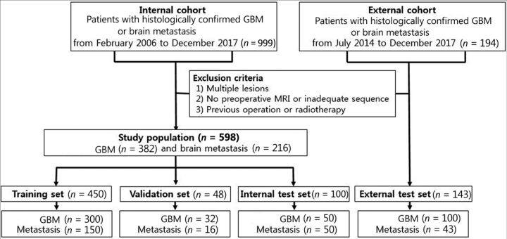
Materials and methods: Records of 598 patients with histologically confirmed glioblastoma or solitary brain metastasis at our institution between February 2006 and December 2017 were retrospectively reviewed. Preoperative contrast-enhanced T1WI and T2WI were preprocessed and roughly segmented with rectangular regions of interest. A deep neural network was trained and validated using MR images from 498 patients. The MR images of the remaining 100 were used as an internal test set. An additional 143 patients from another tertiary hospital were used as an external test set. The classifications of ResNet-50 and 2 neuroradiologists were compared for their accuracy, precision, recall, F1 score, and area under the curve.
Results: The areas under the curve of ResNet-50 were 0.889 and 0.835 in the internal and external test sets, respectively. The area under the curve of neuroradiologists 1 and 2 were 0.889 and 0.768 in the internal test set and 0.857 and 0.708 in the external test set, respectively.
Conclusions: A deep learning-based model may be a supportive tool for preoperative discrimination between glioblastoma and solitary brain metastasis using conventional MR images.
© 2021 by American Journal of Neuroradiology.
Figures


Similar articles
-
Multiple deep learning models based on MRI images in discriminating glioblastoma from solitary brain metastases: a multicentre study.BMC Med Imaging. 2025 May 19;25(1):171. doi: 10.1186/s12880-025-01703-3. BMC Med Imaging. 2025. PMID: 40389875 Free PMC article.
-
Discrimination Between Glioblastoma and Solitary Brain Metastasis Using Conventional MRI and Diffusion-Weighted Imaging Based on a Deep Learning Algorithm.J Digit Imaging. 2023 Aug;36(4):1480-1488. doi: 10.1007/s10278-023-00838-5. Epub 2023 May 8. J Digit Imaging. 2023. PMID: 37156977 Free PMC article.
-
Ensemble learning-based radiomics model for discriminating brain metastasis from glioblastoma.Eur J Radiol. 2025 Feb;183:111900. doi: 10.1016/j.ejrad.2024.111900. Epub 2024 Dec 24. Eur J Radiol. 2025. PMID: 39733718
-
Deep Learning AI Applications in the Imaging of Glioma.Top Magn Reson Imaging. 2020 Apr;29(2):115-0. doi: 10.1097/RMR.0000000000000237. Top Magn Reson Imaging. 2020. PMID: 32271288 Review.
-
Updates on Deep Learning and Glioma: Use of Convolutional Neural Networks to Image Glioma Heterogeneity.Neuroimaging Clin N Am. 2020 Nov;30(4):493-503. doi: 10.1016/j.nic.2020.07.002. Epub 2020 Sep 18. Neuroimaging Clin N Am. 2020. PMID: 33038999 Review.
Cited by
-
Inherited genetics of adult diffuse glioma and polygenic risk scores-a review.Neurooncol Pract. 2022 Mar 12;9(4):259-270. doi: 10.1093/nop/npac017. eCollection 2022 Aug. Neurooncol Pract. 2022. PMID: 35859544 Free PMC article. Review.
-
Aided Diagnosis Model Based on Deep Learning for Glioblastoma, Solitary Brain Metastases, and Primary Central Nervous System Lymphoma with Multi-Modal MRI.Biology (Basel). 2024 Feb 5;13(2):99. doi: 10.3390/biology13020099. Biology (Basel). 2024. PMID: 38392317 Free PMC article.
-
Artificial Intelligence in CT and MR Imaging for Oncological Applications.Cancers (Basel). 2023 Apr 30;15(9):2573. doi: 10.3390/cancers15092573. Cancers (Basel). 2023. PMID: 37174039 Free PMC article. Review.
-
Deep learning models for rapid discrimination of high-grade gliomas from solitary brain metastases using multi-plane T1-weighted contrast-enhanced (T1CE) images.Quant Imaging Med Surg. 2024 Aug 1;14(8):5762-5773. doi: 10.21037/qims-24-380. Epub 2024 Jul 16. Quant Imaging Med Surg. 2024. PMID: 39144024 Free PMC article.
-
Neuroinflammation and immunoregulation in glioblastoma and brain metastases: Recent developments in imaging approaches.Clin Exp Immunol. 2021 Dec;206(3):314-324. doi: 10.1111/cei.13668. Epub 2021 Oct 8. Clin Exp Immunol. 2021. PMID: 34591980 Free PMC article. Review.
References
Publication types
MeSH terms
LinkOut - more resources
Full Text Sources
Other Literature Sources
Medical