. 2021 Mar 18;11(1):6344.
doi: 10.1038/s41598-021-85010-1.

Retinal ganglion cell dysfunction in preclinical Alzheimer's disease: an electrophysiologic biomarker signature

Affiliations

Retinal ganglion cell dysfunction in preclinical Alzheimer's disease: an electrophysiologic biomarker signature

Samuel Asanad et al. Sci Rep. .

Abstract

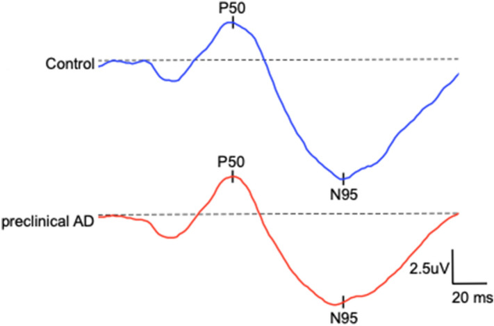
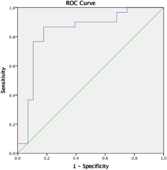
The current study evaluated retinal function using electroretinography (ERG) in cognitively healthy (CH) participants with preclinical Alzheimer's disease (AD), as classified by cerebral spinal fluid (CSF) Aβ42/Tau ratio. Individuals with normal retinal morphology ascertained by spectral-domain optical coherence tomography were enrolled. Full-field ERG, pattern PERG, and photopic negative response (PhNR) were performed in 29 adult participants (58 eyes). Amplitude and implicit times of the ERG wave components were analyzed. Preclinical AD participants showed marked retinal ganglion cell dysfunction relative to controls. The PhNR was significantly diminished in preclinical AD relative to controls. PhNR amplitude and N95 implicit time differentiated CH individuals with CSF biomarkers of AD pathology with 87% sensitivity and 82% specificity. These quantitative electrophysiologic findings expand our understanding of early retinal functional changes that precede cognitive decline in AD. Retinal ganglion cell dysfunction, as detected by ERG, may be a clinically useful, non-invasive in vivo biomarker for early disease detection, which is necessary for ultimately pursuing early intervention.


Conflict of interest statement

The authors declare no competing interests.

Figures

Figure 1
Figure 1
Depicts the recorded pattern electroretinogram responses in representative preclinical Alzheimer’s disease (AD) (mean N95 ± SD: 5.7 ± 2.3 µV) and control participants (mean N95 ± SD: 6.1 ± 1.7 µV).
Figure 2
Figure 2
Comparison of the recorded amplitudes (top) and implicit times (bottom) pattern electroretinogram responses between CH-NAT and CH-PAT participants. CH-NAT, cognitively healthy with normal amyloid/tau ratio; CH-PAT, cognitively healthy with pathologic amyloid/tau ratio. Error bars denote standard error.
Figure 3
Figure 3
Depicts the recorded photopic negative responses (PhNR) in representative preclinical Alzheimer’s disease (AD) and control participants. As illustrated by the double-headed arrow, the PhNR was markedly diminished in preclinical AD as compared to controls.
Figure 4
Figure 4
Photopic negative response (PhNR) comparison between CH-NAT and CH-PAT participants. CH-NAT, cognitively healthy with normal amyloid/tau ratio; CH-PAT, cognitively healthy with pathologic amyloid/tau ratio. Error bars denote standard error, * p < 0.05.
Figure 5
Figure 5
Representation of the ROC curve of the regression analysis to demonstrate which parameters of ERG predict CH-NAT versus CH-PAT group classification. The area under the curve (AUC) of PhNR amplitude and N95 implicit time was 0.84.

References

    1. Alzheimer’s Association Alzheimer’s disease facts and figures. Alzheimers Dement. 2020;16:391–460. doi: 10.1002/alz.12068. - DOI - PubMed
    1. Batsch NL, Mittelman MS. World Alzheimer Report 2012: Overcoming the Stigma of Dementia. London: Alzheimer’s Disease International; 2012.
    1. McKhann G, et al. Clinical diagnosis of Alzheimer’s disease: Report of the NINCDS-ADRDA Work Group* under the auspices of Department of Health and Human Services Task Force on Alzheimer’s Disease. Neurology. 1984;34:939–939. doi: 10.1212/WNL.34.7.939. - DOI - PubMed
    1. Katz B, Rimmer S. Ophthalmologic manifestations of Alzheimer’s disease. Surv. Ophthalmol. 1989;34:31–43. doi: 10.1016/0039-6257(89)90127-6. - DOI - PubMed
    1. Tzekov R, Mullan M. Vision function abnormalities in Alzheimer disease. Surv. Ophthalmol. 2014;59:414–433. doi: 10.1016/j.survophthal.2013.10.002. - DOI - PubMed

Publication types

MeSH terms