. 2021 Mar 18;11(1):6194.
doi: 10.1038/s41598-021-85096-7.

Expression profile of the matricellular protein periostin in paediatric inflammatory bowel disease

Affiliations

Expression profile of the matricellular protein periostin in paediatric inflammatory bowel disease

Tracy Coelho et al. Sci Rep. .

Abstract

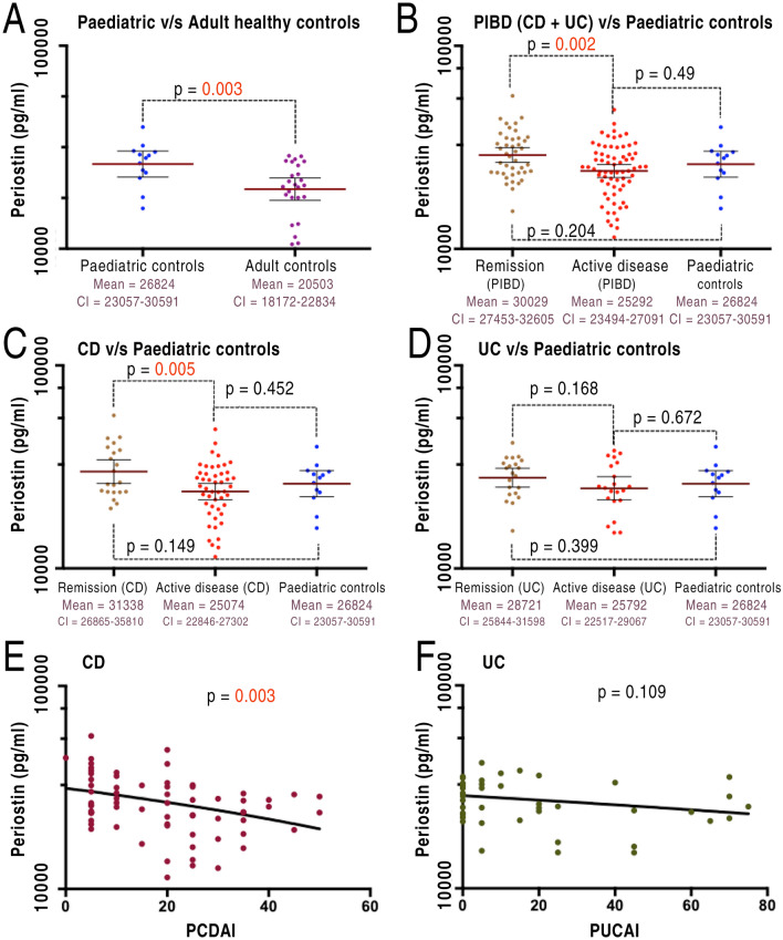
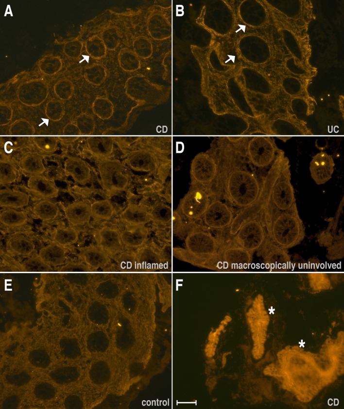
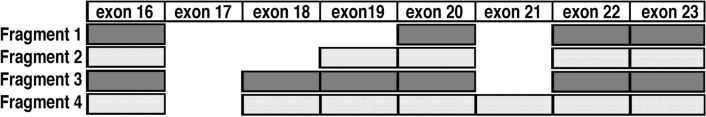
The precise role of periostin, an extra-cellular matrix protein, in inflammatory bowel disease (IBD) is unclear. Here, we investigated periostin in paediatric IBD including its relationship with disease activity, clinical outcomes, genomic variation and expression in the colonic tissue. Plasma periostin was analysed using ELISA in 144 paediatric patients and 38 controls. Plasma levels were assessed against validated disease activity indices in IBD and clinical outcomes. An immuno-fluorescence for periostin and detailed isoform-expression analysis in the colonic tissue was performed in 23 individuals. We integrated a whole-gene based burden metric 'GenePy' to assess the impact of variation in POSTN and 23 other genes functionally connected to periostin. We found that plasma periostin levels were significantly increased during remission compared to active Crohn's disease. The immuno-fluorescence analysis demonstrated enhanced peri-cryptal ring patterns in patients compared to controls, present throughout inflamed, as well as macroscopically non-inflamed colonic tissue. Interestingly, the pattern of isoforms remained unchanged during bowel inflammation compared to healthy controls. In addition to its role during the inflammatory processes in IBD, periostin may have an additional prominent role in mucosal repair. Additional studies will be necessary to understand its role in the pathogenesis, repair and fibrosis in IBD.


Conflict of interest statement

The authors declare no competing interests.

Figures

Figure 1
Figure 1
Plasma periostin levels against disease activity. (A) Significantly lower levels in adult controls in comparison to paediatric controls (p = 0.003). (B) Higher levels during remission (combined for both CD & UC) than active disease (p = 0.002). (C) Statistically significant differences in periostin levels during remission driven by CD; remission (31,338 ± 2144) compared to active disease (25,074 ± 1108); p = 0.005. (D) Patients with UC show no significant differences during remission compared to active disease. (E) An inverse relationship between periostin levels and PCDAI scores (F) No relationship between periostin and disease activity in UC. This figure was, as also mentioned in the methods/stats section, generated through PRISM https://www.graphpad.com/scientific-software/prism/ Version 7. Abbreviations: CD- Crohn’s disease; PCDAI- paediatric Crohn’s disease activity index; PUCAI- Paediatric ulcerative colitis activity index; pIBD- paediatric inflammatory bowel disease (includes both Crohn’s disease and ulcerative colitis); UC- ulcerative colitis.
Figure 2
Figure 2
Immuno-fluorescence staining for periostin in colonic tissue. The figure shows representative immuno-fluorescence for periostin on the colonic tissue obtained from controls and IBD patients in the active state. Note the distinctly localised peri-cryptal rings that can be seen in CD (A, C, D) and UC (B) patients but not in the healthy control tissue (E). Interestingly, the peri-cryptal rings can be seen also in the macroscopically unaffected but histologically inflamed colonic mucosa in CD patients (D). These high intense areas appear more pronounced in CD patients compared to UC patients. Also note the very intense and diffuse staining of the epithelium and stromal tissue in the surgically resected bowel segment of a CD patient (F). Scale bar is 50 µm.
Figure 3
Figure 3
Periostin isoforms in the colonic tissue. The gel shows the isoforms present in tissue biopsies of CD patients (lanes 1–5), UC patient (lanes 6–7) and non-IBD controls (lanes 8–11). Lane 12 shows the negative control. Please note that there are minor fluctuations between the intensities of the different fragments that do not show any correlation between diseases CD (1–5) or UC (6–7), or controls (8–11). Fragment 1 (188 bp) is just below the 200 bp marker while fragment 4 (452 bp) is between 400 and 500 bp.
Figure 4
Figure 4
Sequencing of the periostin isoforms. Based on sequencing the exon usage of the four periostin isoforms isolated from the GI tissue in patients and healthy controls is shown. No alternative splicing occurs in exons 1–16 and the last exon 23 with the stop codon is also present in all variants. Fragment 1 contains exons 16, 20, 22 and 23 and is exactly 188 bp long, while fragment 2 contains additionally exon 19 and is 278 bp long. Fragment 3 additionally has exon 18 present and is 368 bp long and fragment 4 contains also exon 21 and is 452 bp long. Interestingly, we did not identify any trace of the full-length fragment that also retains exon 21 and would be expected to be 536 bp in length.
Figure 5
Figure 5
Network of genes for periostin. Screenshot from PathCards displaying seventeen SuperPath genes (black text) functionally connected to periostin and ten additional genes, known to be related to or influencing periostin signalling (red text). The protein nodes and the inter-connections within the retrieved network are automatically coloured for visual clarity and also to indicate the type of functional connectivity. The genes displayed in this figure are: C-X-C Motif Chemokine Receptor 4 (CXCR4); Cytohesin 1 Interacting Protein (CYTIP); Endothelial PAS Domain Protein 1 (EPAS1); Jagged Canonical Notch Ligand 1 (JAG1); Lymphoid Enhancer Binding Factor 1 (LEF1); Periostin (POSTN); Phosphatidylinositol-4,5-Bisphosphate 3-Kinase Catalytic Subunit Gamma (PIK3CG); SRC Proto-Oncogene, Non-Receptor Tyrosine Kinase (SRC); Tenascin C (TNC); Transcription Factor 7 (TCF7); Transcription Factor 7 Like 1 (TCF7L1); Transcription Factor 7 Like 2 (TCF7L2); Vascular Cell Adhesion Molecule 1 (VCAM1); Von Hippel-Lindau Tumor Suppressor (VHL); Wnt Family Member 2 (WNT2), Rho-associated protein kinase 1 (ROCK1), Ras Homolog Family Member A (RHOA), C–C chemokine receptor type 5 (CCR5), C–C Motif Chemokine Ligand 5 (CCL5), Matrix metalloproteinase-2 (MMP2), Matrix metalloproteinase-9 (MMP9) Signal transducer and activator of transcription 3 (STAT3), Interleukin 6 (IL-6) Integrin alpha V beta 1 & 3 (ITGA5B1&3). The screenshot was taken from the following url in April 2018. https://pathcards.genecards.org/Card/amplification_and_expansion_of_oncogenic_pathways_as_metastatic_traits?queryString=POSTN The screenshot database is based on a publication from Belinky et al. and was modified based on results in recent publications,.
Figure 6
Figure 6
Pathophysiology of periostin. In the tissue, periostin is primarily produced by the fibroblasts and myofibroblasts. It has also been detected in tumour cells, epithelial cells and leukocytes. After engaging with the αv-integrin receptors within the cell membrane, periostin facilitates transmission of signals for cellular adhesion and activation of various immune-signalling pathways. These include the focal adhesion kinase (FAK) phosphatidylinositol 3-kinase (PI3K)/AKT, NF-κB, and RHOA and YAP/TAZ TEAD signalling pathways. These pathways further fuel inflammation and also contribute to tissue repair and remodelling as well as fibrosis and tumorigenesis. This figure was generated by TC using the software https://app.diagrams.net. (Abbreviations: TAZ, transcriptional co-activator with PDZ-binding motif; TEAD, TEA domain family member 1; YAP, Yes-associated protein1).

References

    1. Murphy-Ullrich JE, Sage EH. Revisiting the matricellular concept. Matrix Biol. 2014;37:1–14. doi: 10.1016/j.matbio.2014.07.005. - DOI - PMC - PubMed
    1. Conway SJ, et al. The role of periostin in tissue remodeling across health and disease. Cell. Mol. Life Sci. 2014;71:1279–1288. doi: 10.1007/s00018-013-1494-y. - DOI - PMC - PubMed
    1. Liu AY, Zheng H, Ouyang G. Periostin, a multifunctional matricellular protein in inflammatory and tumor microenvironments. Matrix Biol. 2014;37:150–156. doi: 10.1016/j.matbio.2014.04.007. - DOI - PubMed
    1. Bao S, et al. Periostin potently promotes metastatic growth of colon cancer by augmenting cell survival via the Akt/PKB pathway. Cancer Cell. 2004;5:329–339. doi: 10.1016/S1535-6108(04)00081-9. - DOI - PubMed
    1. Gonzalez-Gonzalez L, Alonso J. Periostin: A matricellular protein with multiple functions in cancer development and progression. Front. Oncol. 2018;8:225. doi: 10.3389/fonc.2018.00225. - DOI - PMC - PubMed

Publication types

MeSH terms