

doi: 10.1038/s41598-021-85425-w.
UV-C irradiation is highly effective in inactivating SARS-CoV-2 replication
Affiliations
- PMID: 33737536
- PMCID: PMC7973506
- DOI: 10.1038/s41598-021-85425-w
Item in Clipboard
UV-C irradiation is highly effective in inactivating SARS-CoV-2 replication
Sci Rep.
.
Abstract
The potential virucidal effects of UV-C irradiation on SARS-CoV-2 were experimentally evaluated for different illumination doses and virus concentrations (1000, 5, 0.05 MOI). At a virus density comparable to that observed in SARS-CoV-2 infection, an UV-C dose of just 3.7 mJ/cm2 was sufficient to achieve a more than 3-log inactivation without any sign of viral replication. Moreover, a complete inactivation at all viral concentrations was observed with 16.9 mJ/cm2. These results could explain the epidemiological trends of COVID-19 and are important for the development of novel sterilizing methods to contain SARS-CoV-2 infection.
Conflict of interest statement
The authors declare no competing interests.
Figures
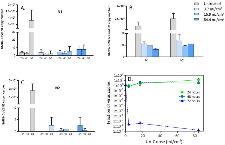
Viral replication of UV-irradiated SARS-CoV-2 (0.05 MOI) virus in in vitro VeroE6 cells. Vero E6 cells were infected with UV-C irradiated SARS-CoV-2 virus at a MOI of 0.05. Culture supernatants were harvested at the indicated times (24, 48 h and 6 days) and virus titers were measured (A,B) by absolute copy number quantification (Real-Time PCR). Viral replication was assessed even on cell lysate harvested at the end of cell cultures (6 days) (C). All cell culture conditions were seeded in duplicate. Panel (D) reports the plots of the measured virus copies normalized at the untreated sample in the different conditions. For descriptive purposes, mean values and whiskers representing the observed half-ranges are shown.

Viral replication of UV-irradiated SARS-CoV-2 (5 MOI) virus in in vitro VeroE6 cells. Vero E6 cells were infected with UV-C irradiated SARS-CoV-2 virus at a MOI of 5. Culture supernatants were harvested at the indicated times (24, 48 and 72 h) and virus titers were measured by absolute copy number quantification (Real-Time PCR, A,B). Viral replication was assessed even on cell lysate harvested at the end of cell cultures (72 h) (C). All cell culture conditions were seeded in duplicate. Panel (D) reports the plots of the measured virus copies normalized at the untreated sample in the different conditions. For descriptive purposes, mean values and whiskers representing the observed half-ranges are shown.

Analyses of virus induced cytopathic effect. (A) No cytopathic effect was observed in uninfected cultured VeroE6 monolayers maintained in 50 mJ/cm2 UV-treated complete medium for 72 h. (B) In vitro infection of SARS-CoV-2 (5 MOI) UV-C untreated VeroE6 cells resulted in an evident cytopathic effect. (C) SARS-CoV-2 irradiation with 3.7 mJ/cm2 UV-C rescued the cytopathic effect induced by UV-C untreated virus.

Viral replication of UV-irradiated SARS-CoV-2 (1000 MOI) virus in in vitro VeroE6 cells. Vero E6 cells were infected with UV-C irradiated SARS-CoV-2 virus at a MOI of 1000. Culture supernatants were harvested at the indicated times (24, 48 and 72 h) and virus titers were measured (A,B) by absolute copy number quantification (Real-Time PCR). Viral replication was assessed even on cell lysate harvested at the end of cell cultures (72 h) (C). All cell culture conditions were seeded in duplicate. Panel (D) reports the plots of the measured virus copies normalized at the untreated sample in the different conditions. For descriptive purposes, mean values and whiskers representing the observed half-ranges are shown.

Mercury lamp spectrum measured in the five positions (A). Inset: scheme of the illuminated dwell and the measuring position. UV–vis transmission spectrum of the Dulbecco’s Modified Eagle’s Medium (DMEM) in a 1 mm quartz cuvette (B).
References
Publication types
MeSH terms
LinkOut - more resources
Full Text Sources
Other Literature Sources
Miscellaneous
