. 2021 Mar 18;11(1):6339.
doi: 10.1038/s41598-021-85577-9.

Differential role of SIRT1/MAPK pathway during cerebral ischemia in rats and humans

Affiliations

Differential role of SIRT1/MAPK pathway during cerebral ischemia in rats and humans

Sireesh Kumar Teertam et al. Sci Rep. .

Abstract

Cerebral ischemia (CI) is a severe cause of neurological dysfunction and mortality. Sirtuin-1 (Silent information regulator family protein 1, SIRT1), an oxidized nicotinamide adenine dinucleotide (NAD+)-dependent protein deacetylase, plays an important role in protection against several neurodegenerative disorders. The present study aims to investigate the protective role of SIRT1 after CI in experimental young and aged rats and humans. Also, the study examines the possible regulatory mechanisms of neuronal death in CI settings. Immunoblotting and immunohistochemistry were used to evaluate changes in the expression of SIRT1, JNK/ERK/MAPK/AKT signaling, and pro-apoptotic caspase-3 in experimental rats and CI patients. The study findings demonstrated that, in aged experimental rats, SIRT1 activation positively influenced JNK and ERK phosphorylation and modulated neuronal survival in AKT-dependent manner. Further, the protection conferred by SIRT1 was effectively reversed by JNK inhibition and increased pro-apoptotic caspase-3 expression. In young experimental rats, SIRT1 activation decreased the phosphorylation of stress-induced JNK, ERK, caspase-3, and increased the phosphorylation of AKT after CI. Inhibition of SIRT1 reversed the protective effect of resveratrol. More importantly, in human patients, SIRT1 expression, phosphorylation of JNK/ERK/MAPK/AKT signaling and caspase-3 were up-regulated. In conclusion, SIRT1 could possibly be involved in the modulation of JNK/ERK/MAPK/AKT signaling pathway in experimental rats and humans after CI.


Conflict of interest statement

The authors declare no competing interests.

Figures

Figure 1
Figure 1
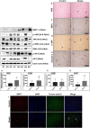
Effect of ischemia on the expression of SIRT1, JNK/ERK/MAPK/AKT signaling pathway in brains of control, 24 h MCAO, MCAO + EX-527, MCAO + resveratrol, and MCAO + SP600125 treatment in young and aged rat. Respective total proteins and beta-tubulin were served as the loading control (p-JNK, p-MEK, p-ERK, and p-AKT), while SIRT1 and p-IRS-1 were normalized to beta-tubulin. (A–F) Statistical results on the changes in expression of SIRT1, p-JNK, p-MEK, p-ERK, p-IRS-1, and p-AKT expression following CI. (G–L) Statistical results on the expression of SIRT1, p-JNK, p-MEK, p-ERK, p-IRS-1, and p-AKT on resveratrol treatment. (M–R) statistical results on the expression of SIRT1, p-JNK, p-MEK, p-ERK, p-IRS-1, and p-AKT on SIRT1 inhibition with EX-527. (S–X) Statistical results on the expression of SIRT1, p-JNK, p-MEK, p-ERK, p-IRS-1, and p-AKT on JNK inhibition with SP600125 treatment. The densitometry values are represented as mean ± SD (n = 5). *p < 0.05 versus sham control.
Figure 2
Figure 2
Assessment of morphological changes following CI. The morphological changes in cortex and hippocampus neurons were assessed using H&E staining (magnification 400X; Scale Bar—50 μm). (A) Representative H&E staining of paraffin-embedded brain sections from sham control, 24 h MCAO, MCAO + resveratrol, MCAO + EX-527, and MCAO + SP600125 treatment in young rats. Morphological changes were represented in black arrows (pyknotic neuron). Black arrow shows the presence of extensive changes in neuronal morphology in the brains of 24 h MCAO and EX-527 treatment, whereas pathological changes was decreased in resveratrol and Sp600125 treatment. (B) H&E staining of brain sections from sham control, 24 h MCAO, MCAO + resveratrol, MCAO + EX-527, and MCAO + SP600125 treatment in aged rats. Black arrow represents the presence of extensive changes in neuronal morphology in the brains of 24 h MCAO and EX-527 treatment, whereas SP600125 and resveratrol failed to reduce pathological changes. The infarct intensity was measured with TTC staining. (C) Resveratrol and SP600125 treatment decreased infarct volume and abolished the effect of EX-527 in the brains of young rat following MCAO. (D) In aged experimental rats EX-527 and SP600125 treatment increased infarct and resveratrol treatment failed to reduce infarct following MCAO. The data are presented as a percentage of tissue loss (n = 3). *p < 0.05 versus ischemic brain.
Figure 3
Figure 3
Subcellular localization of SIRT1, p-JNK, & caspase-3 in aged rats following CI. Aged rat brains were paraffin embedded and sectioned into 5–10 μm thick slices (n = 4). (A) Subcellular distribution of SIRT1 in the brains of aged rat cortex and hippocampus neurons from sham control as well as brains from 24 h after MCAO, MCAO + EX-527, MCAO + resveratrol, and MCAO + SP600125 treatment. (B) p-JNK immunoreactivity in the brains of aged rat cortex and hippocampus neurons from control as well as brains from 24 h after MCAO, MCAO + EX-527, MCAO + resveratrol, and MCAO + SP600125 treatment. (C) Nuclear immunoreactivity of activated caspase-3 in the brains of aged rat cortex and hippocampus neurons from control as well as brains from 24 h after MCAO, MCAO + EX-527, MCAO + resveratrol, and MCAO + SP600125 treatment (magnification 400X; Scale Bar—50 μm). Immunoreactivity for SIRT1, p-JNK, and caspase-3 was depicted in brown (DAB). The neuronal nucleus is visualized with Hematoxylin counterstain.
Figure 4
Figure 4
Subcellular localization of SIRT1, p-JNK, & caspase-3 in young rats following CI. Young rat brains were paraffin embedded and sectioned into 5–10 μm thick slices (n = 4). (A) Subcellular distribution of SIRT1 in the brains of young rat cortex and hippocampus neurons from sham control as well as brains from 24 h after MCAO, MCAO + EX-527, MCAO + resveratrol, and MCAO + SP600125 treatment. (B) p-JNK immunoreactivity in the brains of young rat cortex and hippocampus neurons from control as well as brains from 24 h after MCAO, MCAO + EX-527, MCAO + resveratrol, and MCAO + SP600125 treatment. (C) Nuclear immunoreactivity of activated caspase-3 in the brains of young rat cortex and hippocampus neurons from control as well as brains from 24 h after MCAO, MCAO + EX-527, MCAO + resveratrol, and MCAO + SP600125 treatment (magnification 400X; Scale Bar—50 μm). Immunoreactivity for SIRT1, p-JNK, and caspase-3 was depicted in brown (DAB). The neuronal nucleus is visualized with Hematoxylin counterstain.
Figure 5
Figure 5
Effect of ischemia on the expression of SIRT1/JNK/ERK/AKT signaling pathway in Post-mortem human brain tissue. An equal amount of total protein sample (50 μg) from Control (n = 3) and Stroke brain (n = 5) were probed by western blots to analyze the expression of SIRT1 and phosphorylation status of p-JNK, p-ERK, and p-AKT. Respective total proteins and beta-actin were served as the loading control (p-JNK, p-ERK, and p-AKT), while SIRT1 was normalized to beta-actin. (A–D) represents an increase in SIRT1, p-JNK, p-ERK, and p-AKT protein expression in human stroke brain compared to respective healthy control. Data represented as mean ± SD. *p < 0.05 versus control brain. Human control (n = 3), as well as stroke brains (n = 4), were paraffin embedded and sectioned into 5-10 μm thick slices. (E) Histopathological changes were observed using H&E staining (magnification ×400; Scale Bar—50 μm). (F,G,H) immunoreactivity of SIRT1, p-JNK, and caspase-3 in control and stroke human brain. Black arrow represents changes in immunoreactivity from respective group. Hematoxylin counterstain used for visualization of neuron nucleus. Immunoreactivity for SIRT1, p-JNK, and caspase-3 was depicted in brown (DAB) (magnification ×400; Scale Bar—50 μm). (I) Triple immunofluorescence labeling was used to probe for SIRT1 (Red), DAPI (Blue), and Fluoro Jade-C (Green) in control (n = 3) and stroke human brains (n = 4). The representative image has shown the number of Fluoro Jade-C positive cells that co-localized with SIRT1 in stroke brain compared to control (white arrows), (Scale Bar—50 μm).
Figure 6
Figure 6
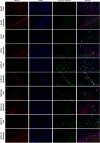
Immunofluorescence labeling of SIRT1 in brains of the aged experimental rat. Triple immunofluorescence staining is used to probe for SIRT1 (Red), DAPI (Blue), and Fluoro Jade-C (Green) to assess neurodegeneration in cortex and hippocampus neurons for control as well as brains from 24 h after MCAO, MCAO + EX-527, MCAO + resveratrol, and MCAO + SP600125 treatment. White arrows indicate co-localization of FJ-C with SIRT1 and DAPI (n = 4; Scale Bar—50 μm).
Figure 7
Figure 7
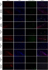
Immunofluorescence labeling of SIRT1 in brains of the young experimental rat. Triple immunofluorescence staining is used to probe for SIRT1 (Red), DAPI (Blue), and Fluoro Jade-C (Green) to assess neurodegeneration in cortex and hippocampus neurons for control as well as brains from 24 h after MCAO, MCAO + EX-527, MCAO + resveratrol, and MCAO + SP600125 treatment. White arrows indicate co-localization of FJ-C with SIRT1 and DAPI (n = 4; Scale Bar—50 μm).

Similar articles

Cited by

References

    1. Shiber JR, Fontane E, Adewale A. Stroke registry: hemorrhagic vs ischemic strokes. Am. J. Emerg. Med. 2010;28:331–333. doi: 10.1016/j.ajem.2008.10.026. - DOI - PubMed
    1. Feigin VL, Norrving B, Mensah GA. Global burden of stroke. Circ. Res. 2017;120:439–448. doi: 10.1161/CIRCRESAHA.116.308413. - DOI - PubMed
    1. Ritzel RM, et al. Aging alters the immunological response to ischemic stroke. Acta Neuropathol. 2018;136:89–110. doi: 10.1007/s00401-018-1859-2. - DOI - PMC - PubMed
    1. Sakamoto J, Miura T, Shimamoto K, Horio Y. Predominant expression of Sir2alpha, an NAD-dependent histone deacetylase, in the embryonic mouse heart and brain. FEBS Lett. 2004;556:281–286. doi: 10.1016/S0014-5793(03)01444-3. - DOI - PubMed
    1. Ramadori G, et al. Brain SIRT1: Anatomical distribution and regulation by energy availability. J. Neurosci. 2008;28:9989–9996. doi: 10.1523/JNEUROSCI.3257-08.2008. - DOI - PMC - PubMed

Publication types