. 2021 Mar 18;11(1):6317.
doi: 10.1038/s41598-021-85613-8.

Karonudib has potent anti-tumor effects in preclinical models of B-cell lymphoma

Affiliations

Karonudib has potent anti-tumor effects in preclinical models of B-cell lymphoma

Morten P Oksvold et al. Sci Rep. .

Abstract

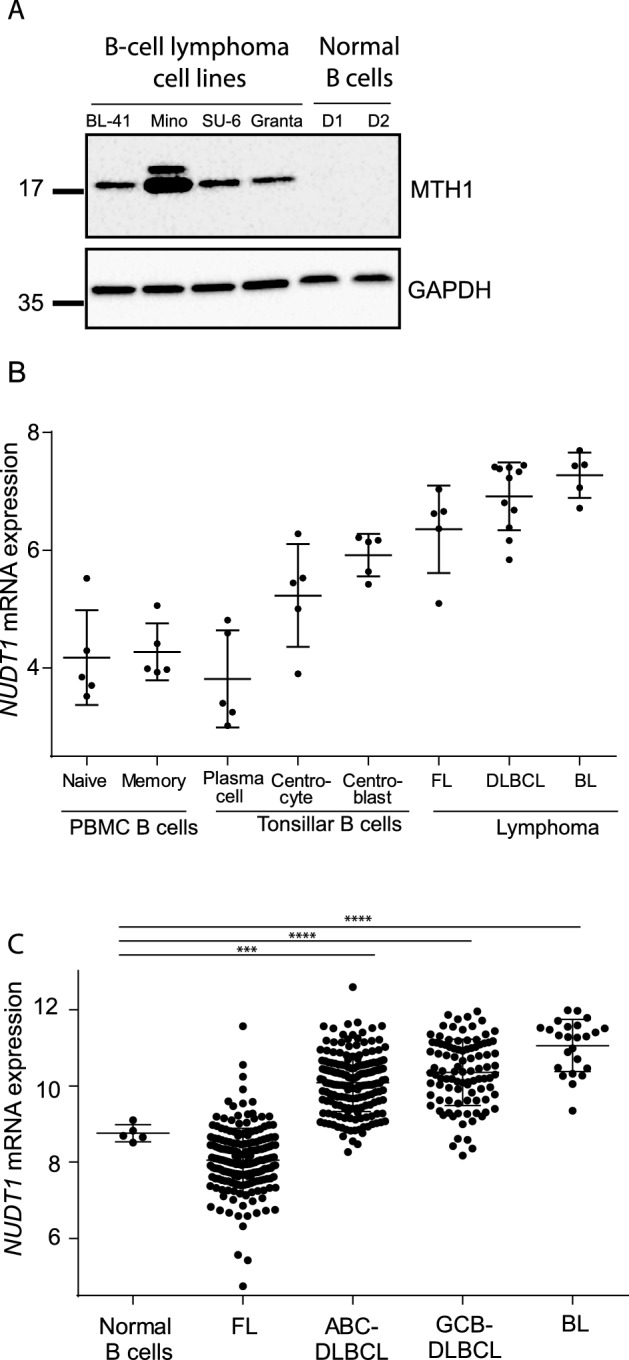
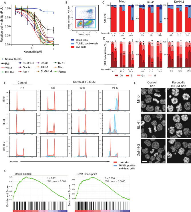
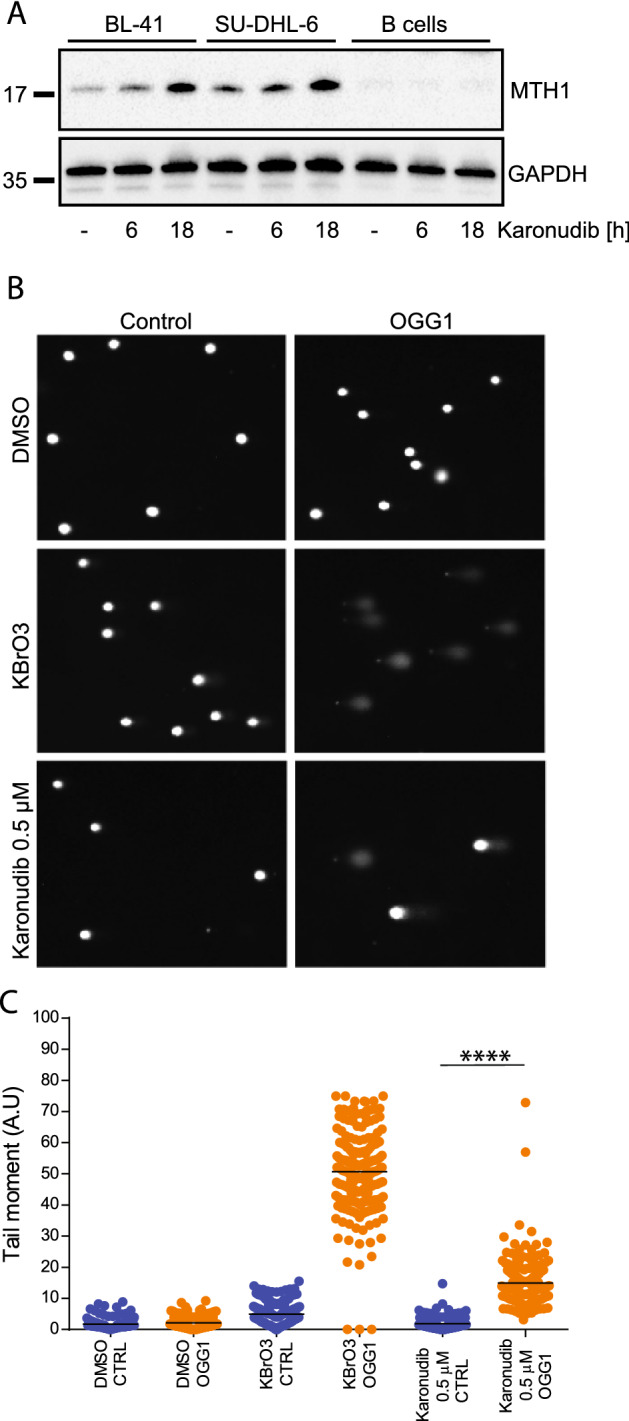
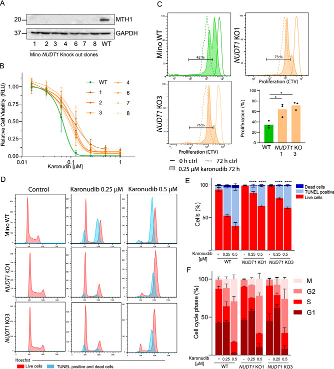
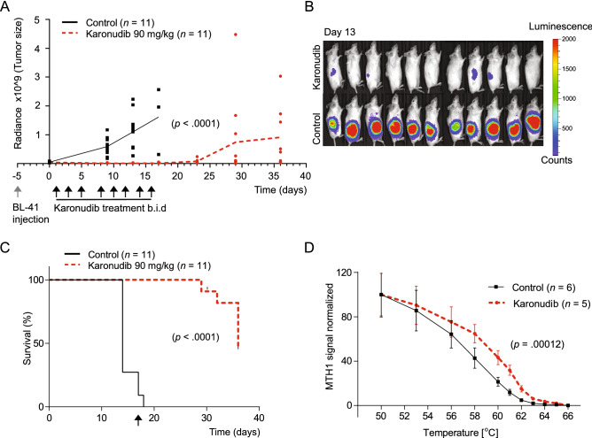
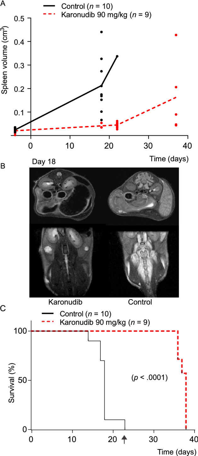
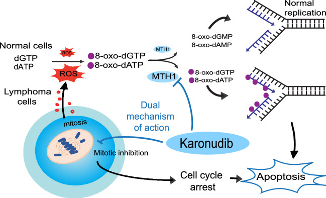
Chemo-immunotherapy has improved survival in B-cell lymphoma patients, but refractory/relapsed diseases still represent a major challenge, urging for development of new therapeutics. Karonudib (TH1579) was developed to inhibit MTH1, an enzyme preventing oxidized dNTP-incorporation in DNA. MTH1 is highly upregulated in tumor biopsies from patients with diffuse large B-cell lymphoma (DLBCL) and Burkitt lymphoma, hence confirming a rationale for targeting MTH1. Here, we tested the efficacy of karonudib in vitro and in preclinical B-cell lymphoma models. Using a range of B-cell lymphoma cell lines, karonudib strongly reduced viability at concentrations well tolerated by activated normal B cells. In B-cell lymphoma cells, karonudib increased incorporation of 8-oxo-dGTP into DNA, and prominently induced prometaphase arrest and apoptosis due to failure in spindle assembly. MTH1 knockout cell lines were less sensitive to karonudib-induced apoptosis, but were displaying cell cycle arrest phenotype similar to the wild type cells, indicating a dual inhibitory role of the drug. Karonudib was highly potent as single agent in two different lymphoma xenograft models, including an ABC DLBCL patient derived xenograft, leading to prolonged survival and fully controlled tumor growth. Together, our preclinical findings provide a rationale for further clinical testing of karonudib in B-cell lymphoma.


Conflict of interest statement

A patent has been filed with TH588 and TH1579 where T.H. is listed as inventor. The Intellectual Property Right is owned by the non-profit Thomas Helleday Foundation for Medical Research (THF). T.H., U.W.B., K.S. and H.G. are board members of the THF. THF is sponsor for on-going clinical trial with TH1579. Oxcia AB is assisting THF in TH1579 clinical trial and U.W.B is chairman of Oxcia AB. H.G., U.W.B., T.P., K.S. and T.H. are shareholders in Oxcia AB. The other authors declare no competing interests.

Figures

Figure 1
Figure 1
MTH1 is upregulated in B-cell lymphoma cell lines and patient samples. (A) MTH1 protein expression in B-lymphoma cell lines and in normal B cells activated with CD40L/IL21 for 24 h (PBMCs, two donors; D1–D2). (B) NUDT1 mRNA data from microdissected samples from subpopulations of B cells from FL (n = 5), BL (n = 5) and DLBCL (n = 11) from Brune et al.32. (C) NUDT1 mRNA levels from the LLMPP-database: FL (n = 191), ABC-DLBCL (n = 176), GCB-DLBCL (n = 97), BL (n = 24), and B cells from PBMCs (n = 5). Stars represent significance compared to normal B cells.
Figure 2
Figure 2
Karonudib induces apoptosis after metaphase arrest in lymphoma cell lines, without affecting healthy B cells. (A) Cell viability was measured by CellTiterGlo and normalized to untreated samples (karonudib, 0.0125–1 µM, 72 h, n = 3). (B) Gating strategy to identify apoptotic cells is shown for Mino cells (karonudib, 0.5 µM, 24 h): TUNEL+, light blue gate;, dead cells, dark blue gate; and live cells in red gate, and (C) the frequencies of these populations in Mino, BL-41 and DoHH-2 (karonudib, 0.5 µM, n = 3). (D) Cell cycle analyses of the live cells (from C). (E) Histograms representing live (red) and dead/apoptotic cells (blue) show G2 accumulation and M-arrest after karonudib treatment. DoHH-2 also show a G1 arrest at 24 h and cells entering apoptosis from G1-arrest. (F) Microscopy of Hoechst stained cells from the same experiment in (BE) (12 h with or without 0.5 µM karonudib, 63×/1.4 objective, bar = 10 µm). An error in the spindle assembly is clearly visible in cells treated with karonudib (images captured randomly), whereas images of successful chromosome segregation in control cells were chosen to illustrate the difference. (G) GSEA of BL-41 and Mino cells (karonudib, 0.5 µM, 12 h), identified Mitotic spindle and G2/M-Checkpoint as significant altered gene sets (P < 0.001 and P = 0.006) (GSEA software v.3.0).
Figure 3
Figure 3
Inhibition of MTH1 leads to increased incorporation of 8-oxo-dGTP. (A) MTH1 expression was detected in total cell lysates (karonudib, 0.5 µM, 6 h, 18 h). (B, C) A modified comet assay was used to indirectly measure incorporation of 8-oxo-dGTP. An increase in comet tails indicates nick in the DNA by recombinant 8-oxoguanin-DNA-glycosylase (OGG-1) where 8-oxo-dGTP is present. BL-41 cells (karonudib, 0.5 µM, 24 h. KBrO3 was used as a positive control (n = 200)). Images are acquired by Zeiss IF microscope and quantified using Comet Assay IV software.
Figure 4
Figure 4
NUDT1 KO in Mino cell lines confirm dual mechanism of karonudib. Two NUDT1 KO clones of Mino were used to assess the effect of MTH1 loss in the lymphoma model. (A) Western immunoblot of seven clones show loss of MTH1 expression. (B) Cell viability was measured in Mino WT (green) and NUDT1 KO (orange) by CellTiterGlo and normalized to untreated samples (karonudib, 0.0125–1 µM, 72 h, n = 3) (C) Proliferation using Cell Trace Violet (CTV) is shown for Mino WT, NUDT1 KO1 and NUDT1 KO3 (continuous line: control cells at time zero, dashed line: control 72 h and filled histogram: 0.25 µM karonudib 72 h). Quantification of the % (shown in gate) of proliferating cells in WT and 0.25 µM karonudib treated cells. *Significant differences (P < 0.05), repeated measurements one-way ANOVA with Dunetts correction between WT and the KO-clones. (D) Gating strategy to identify live, dead and apoptotic cells is shown for Mino cells in Fig. 2B. Histogram representing live (red) and dead/apoptotic cells (blue) show G2 accumulation and M-arrest after karonudib treatment, 24 h. (E) TUNEL+, light blue,; dead cells, dark blue,; and live cells in red show the frequencies of these populations in Mino WT, NUDT1 KO 1 and NUDT1 KO 3 (karonudib, 0.25 and 0.5 µM, 24 h, n = 3). *Significant differences (P < 0.0001, one-way ANOVA with Dunetts correction) between WT and the KO-clones for both concentrations of karonudib. (F) Cell cycle analyses of the live cells from (E).
Figure 5
Figure 5
Karonudib has a strong anti-tumor effect in vivo. (A) Tumor growth of BL-41-luc cells was monitored with IVIS luminescence measurements (physical units of surface radiance (photons/s/cm2/st)) at day 0, 9, 13, 17, 23, 29 and 36 after treatment start. Treatment was initiated 6 days after inoculation, and karonudib was given per os, b.i.d, 90 mg/kg three times a week for 16 days. Tumor growth was significantly decreased in mice treated with karonudib already at day 9, 13 and 17 compared with treatment start (P < 0.0001 for all time points). There was a significant difference (P < 0.0001) between treatment and vehicle both 9 and 13 days after treatment start (unpaired two-tailed t-test with Welsh's correction). (B) IVIS image of animals at day 13. (C) Kaplan–Meier is based on tumor size (> 2 cm3 or 2 cm length in one direction). Median survival was 14 vs. 36 days in control vs. karonudib treated mice (P < 0.0001, Log-rank test). Arrow indicates end of treatment. (D) In vivo target engagement of karonudib to MTH1 shown by CETSA. Vehicle treated mice were randomized and then given karonudib (90 mg/kg) or vehicle 18 h and 4 h prior to euthanization and dissection of tumor tissue. Melting curve for MTH1 with a significant Tm shift of 2.55°C is shown (P = 0.00012, karonudib treated (n = 5), vehicle (n = 6)) measured at 50% MTH1 denaturation.
Figure 6
Figure 6
Karonudib has a strong anti-tumor effect in the ABC DLBCL PDX model DFBL-49659-V2. Tumor cells were injected intravenously and tumor growth monitoring of spleen and bone marrow with MR-imaging. (A) The treatment was initiated 12 days after tumor injection, and continued for 23 days as indicated (karonudib per os, b.i.d, 90 mg/kg three times a week). Spleen volume was measured by manual delineation on the T2-weighted images. First scan was performed 4 days prior to treatment start up. (B) T2-weighted MRI scans of a karonudib and vehicle treated mice at day 18. The arrows indicate increased spleen and edema around the spine in the untreated animal. (C) Median survival was 18 vs. 38 days in control vs. karonudib treated mice (P < 0.0001, Log-rank test). The mice were monitored until they showed clinical signs. Arrow indicates end of treatment. All images are presented in Supplemental Fig. 6A.
Figure 7
Figure 7
Dual mechanism of karonudib leads to apoptosis in lymphoma cells. Karonudib was developed to target the nucleotide metabolism by inhibiting the nucleotide pool sanitizing enzyme, MTH1. MTH1 converts oxidized nucleotide triphosphates created by reactive oxygen species (ROS) to the corresponding monophosphate forms, preventing incorporation of oxidized nucleotides into DNA. High ROS levels in lymphoma corresponds to high MTH1 levels. Karonudib inhibits the function of MTH1 and results in increased oxidized nucleotides in the DNA, and the drug also perturbs microtubule polymerization and leads to cell cycle arrest and apoptosis.

Similar articles

  • MTH1 Inhibitor TH1579 Induces Oxidative DNA Damage and Mitotic Arrest in Acute Myeloid Leukemia.
    Sanjiv K, Calderón-Montaño JM, Pham TM, Erkers T, Tsuber V, Almlöf I, Höglund A, Heshmati Y, Seashore-Ludlow B, Nagesh Danda A, Gad H, Wiita E, Göktürk C, Rasti A, Friedrich S, Centio A, Estruch M, Våtsveen TK, Struyf N, Visnes T, Scobie M, Koolmeister T, Henriksson M, Wallner O, Sandvall T, Lehmann S, Theilgaard-Mönch K, Garnett MJ, Östling P, Walfridsson J, Helleday T, Warpman Berglund U. Sanjiv K, et al. Cancer Res. 2021 Nov 15;81(22):5733-5744. doi: 10.1158/0008-5472.CAN-21-0061. Epub 2021 Sep 30. Cancer Res. 2021. PMID: 34593524 Free PMC article.
  • Validation and development of MTH1 inhibitors for treatment of cancer.
    Warpman Berglund U, Sanjiv K, Gad H, Kalderén C, Koolmeister T, Pham T, Gokturk C, Jafari R, Maddalo G, Seashore-Ludlow B, Chernobrovkin A, Manoilov A, Pateras IS, Rasti A, Jemth AS, Almlöf I, Loseva O, Visnes T, Einarsdottir BO, Gaugaz FZ, Saleh A, Platzack B, Wallner OA, Vallin KS, Henriksson M, Wakchaure P, Borhade S, Herr P, Kallberg Y, Baranczewski P, Homan EJ, Wiita E, Nagpal V, Meijer T, Schipper N, Rudd SG, Bräutigam L, Lindqvist A, Filppula A, Lee TC, Artursson P, Nilsson JA, Gorgoulis VG, Lehtiö J, Zubarev RA, Scobie M, Helleday T. Warpman Berglund U, et al. Ann Oncol. 2016 Dec;27(12):2275-2283. doi: 10.1093/annonc/mdw429. Epub 2016 Nov 8. Ann Oncol. 2016. PMID: 27827301
  • AXL and CAV-1 play a role for MTH1 inhibitor TH1579 sensitivity in cutaneous malignant melanoma.
    Das I, Gad H, Bräutigam L, Pudelko L, Tuominen R, Höiom V, Almlöf I, Rajagopal V, Hansson J, Helleday T, Egyházi Brage S, Warpman Berglund U. Das I, et al. Cell Death Differ. 2020 Jul;27(7):2081-2098. doi: 10.1038/s41418-019-0488-1. Epub 2020 Jan 9. Cell Death Differ. 2020. PMID: 31919461 Free PMC article.
  • TH1579, MTH1 inhibitor, delays tumour growth and inhibits metastases development in osteosarcoma model.
    Moukengue B, Brown HK, Charrier C, Battaglia S, Baud'huin M, Quillard T, Pham TM, Pateras IS, Gorgoulis VG, Helleday T, Heymann D, Berglund UW, Ory B, Lamoureux F. Moukengue B, et al. EBioMedicine. 2020 Mar;53:102704. doi: 10.1016/j.ebiom.2020.102704. Epub 2020 Mar 7. EBioMedicine. 2020. PMID: 32151797 Free PMC article.
  • Potent and specific MTH1 inhibitors targeting gastric cancer.
    Zhou W, Ma L, Yang J, Qiao H, Li L, Guo Q, Ma J, Zhao L, Wang J, Jiang G, Wan X, Adam Goscinski M, Ding L, Zheng Y, Li W, Liu H, Suo Z, Zhao W. Zhou W, et al. Cell Death Dis. 2019 Jun 4;10(6):434. doi: 10.1038/s41419-019-1665-3. Cell Death Dis. 2019. PMID: 31164636 Free PMC article.

Cited by

References

    1. Coiffier B, Sarkozy C. Diffuse large B-cell lymphoma: R-CHOP failure-what to do? Hematol. Am. Soc. Hematol. Educ. Program. 2016;366–378:2016. doi: 10.1182/asheducation-2016.1.366. - DOI - PMC - PubMed
    1. Gisselbrecht C, et al. Salvage regimens with autologous transplantation for relapsed large B-cell lymphoma in the rituximab era. J. Clin. Oncol. 2010;28:4184–4190. doi: 10.1200/JCO.2010.28.1618. - DOI - PMC - PubMed
    1. Casulo C, Friedberg JW. Burkitt lymphoma—A rare but challenging lymphoma. Best Pract. Res. Clin. Haematol. 2018;31:279–284. doi: 10.1016/j.beha.2018.07.013. - DOI - PubMed
    1. Maddocks K. Update on mantle cell lymphoma. Blood. 2018;132:1647–1656. doi: 10.1182/blood-2018-03-791392. - DOI - PubMed
    1. Berglund UW, et al. Validation and development of MTH1 inhibitors for treatment of cancer. Ann. Oncol. 2016;27:2275–2283. doi: 10.1093/annonc/mdw429. - DOI - PubMed

Publication types

MeSH terms