

doi: 10.1038/s41598-021-85667-8.
Intranuclear birefringent inclusions in paraffin sections by polychromatic polarization microscopy
Affiliations
- PMID: 33737593
- PMCID: PMC7973427
- DOI: 10.1038/s41598-021-85667-8
Item in Clipboard
Intranuclear birefringent inclusions in paraffin sections by polychromatic polarization microscopy
Sci Rep.
.
Abstract
Intranuclear birefringent inclusions (IBI) found in various cell types in paraffin-embedded tissue sections have long been considered to be a tissue processing artifact, although an association with biological processes has been suggested. We applied polychromatic polarization microscopy to image their spatial organization. Our study provides evidence that IBI are caused by liquid paraffin-macromolecular crystals formed during paraffin-embedding procedures within cells and potentially reflect an active transcriptional status.
Conflict of interest statement
The authors declare no competing interests.
Figures
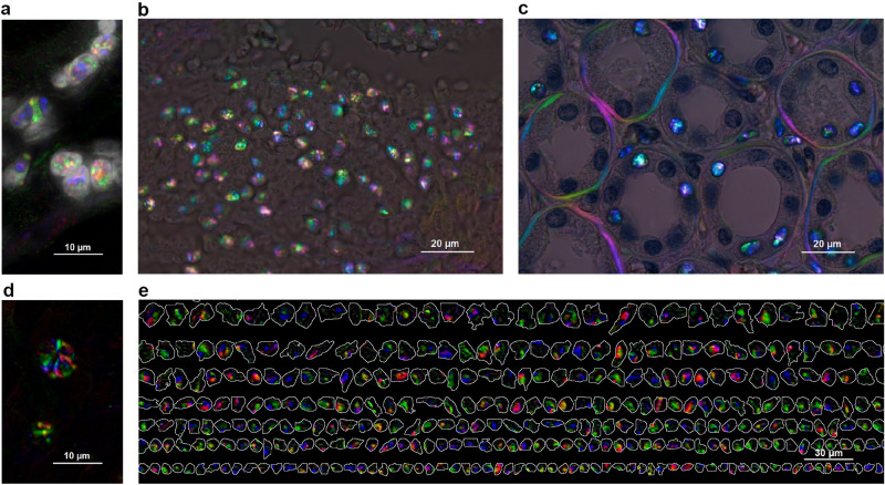
Spatial patterns of IBI captured by PPM. (a) Extracted PPM signals of intranuclear inclusions overlaid with Hoechst-stained fluorescence image for nuclei visualization; (b) endometrioid carcinoma tissue section by PPM after deparaffinization showing a cluster of IBI; (c) IBI and basement membrane collagen PPM signals in kidney tissue section stained with hematoxylin for nuclei visualization and imaged using PPM; (d) Crystalloid structure of the IBI—nuclear inclusions captured in FFPE unstained tissue sections imaged by PPM with digital background correction; (e) gallery of nuclei segmented from Hoechst-stained renal cell carcinoma tissue imaged by fluorescence and overlaid with IBI acquired by PPM. All images acquired with Olympus BX63 microscope equipped with an Olympus DP80 camera using the CellSens acquisition software. Objectives: (b,c) Olympus UPlanApo 100x, (a,d) additional ×2 magnification lens used, (e) Olympus LUCPlanFLN 20x, with Olympus U_FUNA DAPI fluorescence filter cube. Original images acquired at a resolution of 1360 × 1024 px, resized and put into multi-panel figure using the GIMP image editor.
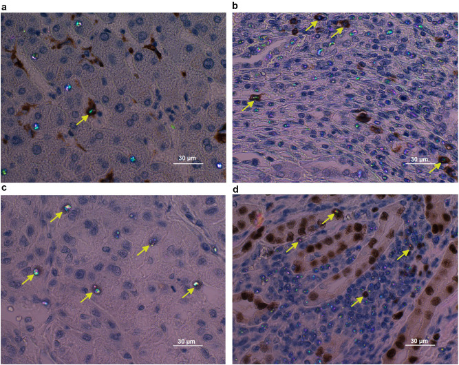
IHC studies to identify IBI-positive cell types and biological processes: (a) IBI present in CD68-positive macrophages (arrow) but also in CD68-negative cells (hepatocellular carcinoma); (b) IBI present in CD20-positive B cells (arrow) but also in CD20-negative cells (renal cell carcinoma); (c) PAX2 marker-specific B cells transcription factor—on renal cell carcinoma tissue section; (d) PAX8 marker-specific renal cancers transcription factor—on renal cell carcinoma tissue section. All images acquired with Olympus BX63 microscope, DP80 camera, objective Olympus Ach 60 × and PPM modality. Original images acquired at a resolution of 1360 × 1024 px, resized and put into multi-panel figure using the GIMP image editor.
Similar articles
-
Evaluation of crystals in formalin-fixed, paraffin-embedded tissue sections for the differential diagnosis of pseudogout, gout, and tumoral calcinosis.Mod Pathol. 2001 Aug;14(8):806-10. doi: 10.1038/modpathol.3880394. Mod Pathol. 2001. PMID: 11504841
-
Analysis of hepcidin expression: in situ hybridization and quantitative polymerase chain reaction from paraffin sections.World J Gastroenterol. 2012 Jul 28;18(28):3727-31. doi: 10.3748/wjg.v18.i28.3727. World J Gastroenterol. 2012. PMID: 22851866 Free PMC article.
-
Simple non-staining method to demonstrate urate crystals in formalin-fixed, paraffin-embedded skin biopsies.J Cutan Pathol. 2009 May;36(5):560-4. doi: 10.1111/j.1600-0560.2008.01116.x. Epub 2009 Mar 9. J Cutan Pathol. 2009. PMID: 19476524
-
Collision and composite tumors; radiologic and pathologic correlation.Abdom Radiol (NY). 2017 Dec;42(12):2909-2926. doi: 10.1007/s00261-017-1200-x. Abdom Radiol (NY). 2017. PMID: 28623377 Review.
-
Complete solubilization of formalin-fixed, paraffin-embedded tissue may improve proteomic studies.Proteomics Clin Appl. 2013 Apr;7(3-4):264-72. doi: 10.1002/prca.201200031. Epub 2013 Mar 6. Proteomics Clin Appl. 2013. PMID: 23339100 Free PMC article. Review.
Cited by
-
Backscattering Mueller matrix polarimetry estimates microscale anisotropy and orientation in complex brain tissue structure.J Med Imaging (Bellingham). 2025 Jan;12(1):016001. doi: 10.1117/1.JMI.12.1.016001. Epub 2024 Dec 31. J Med Imaging (Bellingham). 2025. PMID: 39744151
-
High-fidelity and rapid cellular-level Mueller matrix imaging for tissue identification with unstained sections.Biomed Opt Express. 2021 Jul 12;12(8):4745-4758. doi: 10.1364/BOE.427614. eCollection 2021 Aug 1. Biomed Opt Express. 2021. PMID: 34513222 Free PMC article.
References
-
- Nedzel GA. Intranuclear birefringent inclusions, an artifact occurring in paraffin sections. Q. J. Microsc. Sci. 1951;s3–92(19):343–346.
-
- Kordan HA. Polarized light and phase-contrast studies of stained and unstained plant cell nucleoli. J. Microsc. 1970;92(2):99–111. doi: 10.1111/j.1365-2818.1970.tb02242.x. - DOI
Publication types
MeSH terms
Substances
Grants and funding
LinkOut - more resources
Full Text Sources
Other Literature Sources
Medical
