. 2021 Mar 18;11(1):6217.
doi: 10.1038/s41598-021-85553-3.

Utilization of patterned bioprinting for heterogeneous and physiologically representative reconstructed epidermal skin models

Affiliations

Utilization of patterned bioprinting for heterogeneous and physiologically representative reconstructed epidermal skin models

Sabrina Madiedo-Podvrsan et al. Sci Rep. .

Erratum in

Abstract

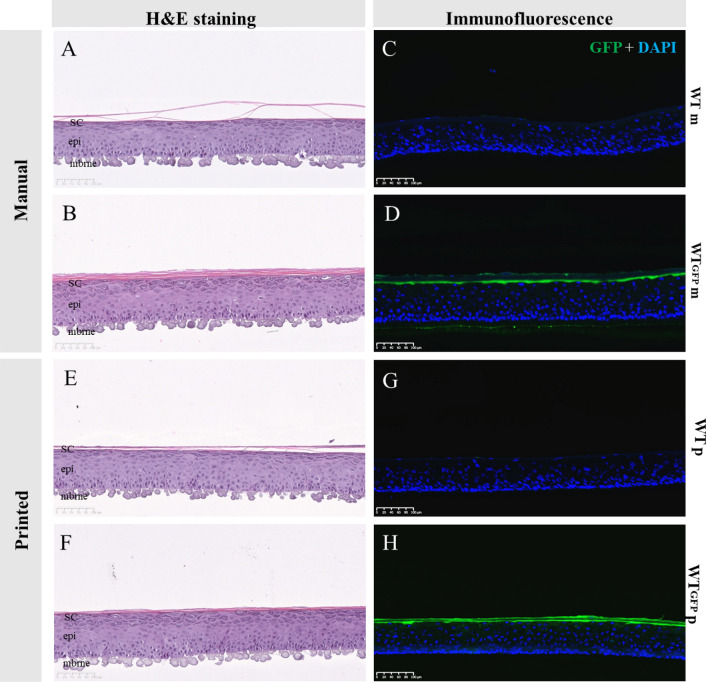
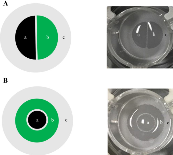
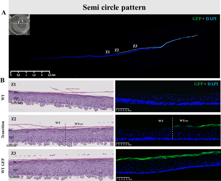
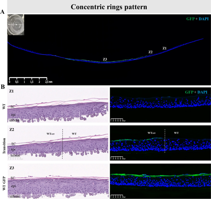
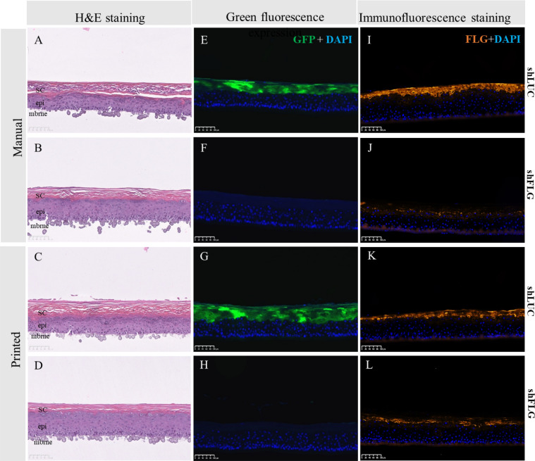
Organotypic skin tissue models have decades of use for basic research applications, the treatment of burns, and for efficacy/safety evaluation studies. The complex and heterogeneous nature of native human skin however creates difficulties for the construction of physiologically comparable organotypic models. Within the present study, we utilized bioprinting technology for the controlled deposition of separate keratinocyte subpopulations to create a reconstructed epidermis with two distinct halves in a single insert, each comprised of a different keratinocyte sub-population, in order to better model heterogonous skin and reduce inter-sample variability. As an initial proof-of-concept, we created a patterned epidermal skin model using GPF positive and negative keratinocyte subpopulations, both printed into 2 halves of a reconstructed skin insert, demonstrating the feasibility of this approach. We then demonstrated the physiological relevance of this bioprinting technique by generating a heterogeneous model comprised of dual keratinocyte population with either normal or low filaggrin expression. The resultant model exhibited a well-organized epidermal structure with each half possessing the phenotypic characteristics of its constituent cells, indicative of a successful and stable tissue reconstruction. This patterned skin model aims to mimic the edge of lesions as seen in atopic dermatitis or ichthyosis vulgaris, while the use of two populations within a single insert allows for paired statistics in evaluation studies, likely increasing study statistical power and reducing the number of models required per study. This is the first report of human patterned epidermal model using a predefined bioprinted designs, and demonstrates the relevance of bioprinting to faithfully reproduce human skin microanatomy.


Conflict of interest statement

Authors declare do not have financial and non-financial interest but declare working in cosmetic company which have financial interest due to cosmetic and tissue engineering business.

Figures

Figure 1
Figure 1
Effect of GFP transfection and bioprinting on epidermal reconstruction morphology. H&E morphology between skin models reconstructed from WT or WT-GFP cells was consistent for both manually (A,B) or bioprinted (E,F) samples. As anticipated, samples printed with WT-GFP (D,H) but not WT cells (C,G) express GPF, with no difference between those using manual (C,D) or (G,H) reconstruction techniques. SC stratum corneum, epi epidermis, mbrne polycarbonate membrane support, WTm manual reconstructed skin substitute from WT NHKs, WTp printed reconstructed skin substitute from WT NHKs, WT-GFPm manual reconstructed skin substitute from WT-GFP NHK, WT-GFPm printed reconstructed skin substitute from WT-GFP NHK (n = 6 for each condition).
Figure 2
Figure 2
Macroscopic visualization of the cellular deposition design. Cells were printed in patterned cellular suspension in either semi-circles (A) or concentric rings (B) schematically illustrated as 2 separate NHK populations (left) or after seeding on a polycarbonate membrane after the bioprinting process (right). Pattern designs were drawn internally using BioCaD software (RegenHU).
Figure 3
Figure 3
Histology of pattern bioprinted epidermis comprised of two distinct cell populations. (A) Lateral view of the reconstructed skin stained for GFP (green) and cell nuclei (DAPI, blue) at day 14 post-reconstruction. Indicated positions Z1–3 correspond to (B) H&E and immunofluorescence stained sections Z1–3, whereby Z1 is from the WT printed cell half, Z2 is the WT/WT-GFP cell interface, and Z3 is representative of the WT-GFP printed half. The H&E images (left) show no difference in gross morphology, while the immunofluorescence images (right) reveal no GFP staining on the WT cell side of the model, a clear demarcation where the WT and WT-GFP halves meet, and a consistent GFP signal in the WT-GFP printed half, demonstrating the model patterning to be stable across 14 days of culture. Images representative of 10 separate models.
Figure 4
Figure 4
Bioprinted RHE constructs were achieved using distinct populations of NHKs on a same sample in order to pattern the concentric design. Morphological analysis of the printed epidermal substitutes and validation of the pattern using H&E staining and fluorescence analysis were done after 14 days of culture at air–liquid interface. (A) GFP staining (Green) on the entire skin substitute with nuclei counterstain with DAPI (blue) where we can observe both conserved parts of the epidermal sample, Z1, Z2 and Z3 are for zone 1, zone 2 and zone 3 represent the areas illustrated in (B); a macroscopic picture of the semi-circle sample has been represented with the orientation of sample slices for the analysis; (B) H&E staining and Green fluorescence analysis on the WT part of the sample (Z1), the transition between the WT and the WTGFP parts (Z2) and the WTGFP part (Z3) proving that pattern is conserved after 14 days of culture and that both NHK populations form at the end of the culture a unique sample. Sc stratum corneum, epi epidermis, mbrne polycarbonate membrane support, WT reconstructed epidermal part from native phenotypic keratinocytes, WTGFP reconstructed skin substitute from GFP-transduced native phenotypic keratinocytes, Transition specific region of the patterned reconstructed skin substitute where both WT and WTGFP are in contact (n = 11).
Figure 5
Figure 5
Histological examination of skin models reconstructed via manual seeding or bioprinting with shLUC or shFLG cells. (A) H&E sections of shLUC reconstructed samples manually seeded show a well-organized epidermal architecture, while (B) shFLG reconstructed samples demonstrate a pronounced hypogranulosis, consistent with FLG down-regulated skin. These data are consistent with those obtained via bioprinting (C,D). Manually seeded shLUC models expressed high levels of (E) GFP, which is not presented in shFLG samples (F). Manually seeded samples are indistinguishable from their bioprinted counterparts (G,H). Lastly, manually printed shLUC models highly express FLG (I), in contrast to samples reconstructed with shFLG NHK (J). There is no discernible difference in expression in models reconstructed using bioprinting (K,J). Images representative of 8 separate samples. SC stratum corneum, epi epidermis, mbrne polycarbonate membrane support.
Figure 6
Figure 6
Expression of FLG and GFP in the reconstructed epidermis created via patterned bioprinting with shLUC and shFLG cell populations. (A) GFP (green) and (B) FLG expression is only present on the shLUC half of the reconstructed skin. Images representative of 8 samples.
Figure 7
Figure 7
Histological analysis and GFP expression in shLUC and shFLG halves of pattern bioprinted reconstructed skin. (A) The suLUC half of the patterned model is histologically normal and shows high levels of GFP staining as anticipated, which abruptly ceases at the border with the shFLG printed half (B). (C) The shFLG printed half of the model demonstrates a lack of keratohyalin granules in H&E staining and no GFP signal, demonstrating the model to be accurately printed and stable with no migration of cell populations after 14 days in culture. Images representative of 8 individual samples.

References

    1. Wong R, Geyer S, Weninger W, Guimberteau JC, Wong JK. The dynamic anatomy and patterning of skin. Exp. Dermatol. 2016;25:92–98. doi: 10.1111/exd.12832. - DOI - PubMed
    1. Oh J, et al. Biogeography and individuality shape function in the human skin metagenome. Nature. 2014;514:59–64. doi: 10.1038/nature13786. - DOI - PMC - PubMed
    1. Fernandez-Flores A. Regional variations in the histology of the skin. Am. J. Dermatopathol. 2015;37:737–754. doi: 10.1097/dad.0000000000000353. - DOI - PubMed
    1. Bell E, Ehrlich HP, Buttle DJ, Nakatsuji T. Living tissue formed in vitro and accepted as skin-equivalent tissue of full thickness. Science (New York, N.Y.) 1981;211:1052–1054. doi: 10.1126/science.7008197. - DOI - PubMed
    1. Albanna M, et al. In situ bioprinting of autologous skin cells accelerates wound healing of extensive excisional full-thickness wounds. Sci. Rep. 2019;9:1856. doi: 10.1038/s41598-018-38366-w. - DOI - PMC - PubMed

MeSH terms