. 2021 Mar 18;11(1):6326.
doi: 10.1038/s41598-021-85866-3.

Curvature sensing amphipathic helix in the C-terminus of RTNLB13 is conserved in all endoplasmic reticulum shaping reticulons in Arabidopsis thaliana

Affiliations

Curvature sensing amphipathic helix in the C-terminus of RTNLB13 is conserved in all endoplasmic reticulum shaping reticulons in Arabidopsis thaliana

Rhiannon L Brooks et al. Sci Rep. .

Abstract

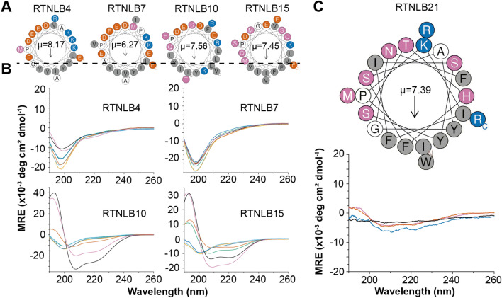
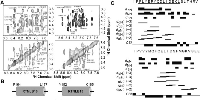
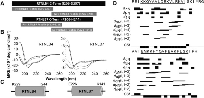
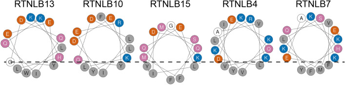
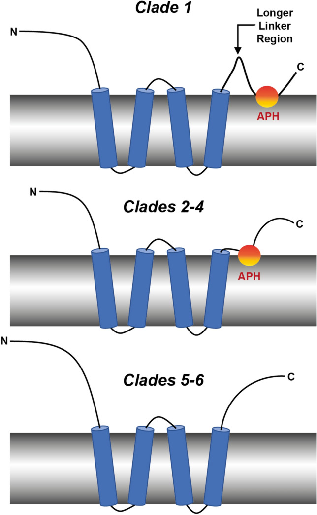
The reticulon family of integral membrane proteins are conserved across all eukaryotes and typically localize to the endoplasmic reticulum (ER), where they are involved in generating highly-curved tubules. We recently demonstrated that Reticulon-like protein B13 (RTNLB13) from Arabidopsis thaliana contains a curvature-responsive amphipathic helix (APH) important for the proteins' ability to induce curvature in the ER membrane, but incapable of generating curvature by itself. We suggested it acts as a feedback element, only folding/binding once a sufficient degree of curvature has been achieved, and stabilizes curvature without disrupting the bilayer. However, it remains unclear whether this is unique to RTNLB13 or is conserved across all reticulons-to date, experimental evidence has only been reported for two reticulons. Here we used biophysical methods to characterize a minimal library of putative APH peptides from across the 21 A. thaliana isoforms. We found that reticulons with the closest evolutionary relationship to RTNLB13 contain curvature-sensing APHs in the same location with sequence conservation. Our data reveal that a more distantly-related branch of reticulons developed a ~ 20-residue linker between the transmembrane domain and APH. This may facilitate functional flexibility as previous studies have linked these isoforms not only to ER remodeling but other cellular activities.


Conflict of interest statement

The authors declare no competing interests.

Figures

Figure 1
Figure 1
(A) Sequence alignment of RTNLB13 APH region of all reticulon-like (RTNLB) protein isoforms found in A. thaliana, listed in order from RTNLB1-RTNLB21. Highly conserved residues are highlighted in black, with the shading of the residues decreasing with decreasing conservation. (B) Sequence logo to indicate the degree of conservation of the RNLB13 APH-region amongst all RTNLB isoforms in A. thaliana. (C) Predicted topologies of representative RTNLB isoforms from each clade. The bar at the top refers to number of amino-acids. Each reticulon contains the RHD in orange and the N-terminus, C-terminus and loop regions in violet. Other regions of functionality for specific RTNs are highlighted in magenta, including the APH in RTNLB13 and the 3 beta-hydroxysteroid-dehydrogenase/decarboxylase domain in RTNLB20. (D) Phylogenetic analysis of all 21 RTNLB isoforms in A. thaliana. The isoforms were then split into six clades based on their evolutionary relationships.
Figure 2
Figure 2
(A) Helical wheel plots of the four representative RTNLB peptides chosen for characterization. Each fits the requirements for an APH with distinct hydrophobic and polar faces, and high hydrophobic moments (μ). Hydrophobic residues are shown in grey, acidic residues in red–orange, basic residues in blue, polar residues in purple and other residues in white. (B) CD spectra of the four peptides (~ 50 µM) in phosphate buffer and a range of model membranes of different diameters. Specifically, measurements were made in DPC micelles (black, 3.7 nm), LMPG micelles (purple, 4.2 nm), DMPC vesicles (blue, 75 nm), DHPC/DMPC bicelles q = 0.25 (red–orange, 18 nm), and 60:1:15 DMPC:DMPG:DHPC vesicles with diameters of 22 nm (green), 42 nm (sky-blue), and 83 nm (orange). All CD data are given in units of mean residue ellipticity (MRE, deg cm2/dmol) and the diameters of the membrane mimetics were estimated using DLS. (C) Helical wheel plot of the RTNLB21 peptide and CD spectra of this peptide (17 µM) in buffer (black), DPC (purple), LMPG (red–orange) and DHPC/DMPC bicelles q = 0.25 (blue).
Figure 3
Figure 3
(A) Representative 1H-1H NOESY NMR data for the RTNLB10 (left) and RTNLB15 (right) peptides (~ 1 mM) solubilized in 50 mM DPC-d38 at a mixing time of 90 ms. The top panels show the fingerprint regions with characteristic α-helical NOEs labelled (i.e. Hα of residue i, NH of residues i + 1, 3, 4). The bottom panels show the region of the spectra where backbone amide NOEs are found, also characteristic for α-helices (i.e. NH of residue i, NH of residues i + 1, 2). (B) Schematic showing the NMR-identified locations of the APH regions in RTNLB10 and RTNLB15. (C) Survey of NMR-derived sequential backbone NOE connectivities for RTNLB10 (Top) and RTNLB15 (Bottom) peptides solubilized in 50 mM DPC-d38, classified as strong, weak, or absent by the thickness (or absence) of a bar connecting the residues involved. The non-sequential connectivities listed (i.e. i, i + n) are unique to helices, and were used alongside chemical shift index analyses (CSI) to localize the amphipathic helices to those residues underlined in the sequences.
Figure 4
Figure 4
(A) Schematic to show the locations of additional RTNLB4 and RTNLB7 peptides further along the C-termini of these two proteins. (B) CD spectra of the RTNLB4 (left) and RTNLB7 (right) peptides (~ 50 µM) in phosphate buffer and a range of model membranes: DPC micelles ( , 3.7 nm), LMPG micelles ( — , 4.2 nm), DMPC vesicles ( · · · , 75 nm), DHPC/DMPC bicelles q = 0.25 (- - -, 18 nm), and 60:1:15 DMPC:DMPG:DHPC vesicles with diameters of 22 nm ( – – – ), 42 nm ( – · – ), and 83 nm (-· ·-). All CD data are given in units of mean residue ellipticity (MRE, deg cm2/dmol) and the diameters of model membranes were estimated using DLS. (C) Schematic showing the NMR-identified locations of the APH regions in RTNLB4 and RTNLB7. (D) Survey of NMR-derived sequential backbone NOE connectivities for RTNLB4 and RTNLB7 peptides solubilized in 50 mM DPC-d38, classified as strong, weak, or absent by the thickness (or absence) of a bar connecting the residues involved. The non-sequential connectivities listed (i.e. i, i + n) are unique to helices, and were used alongside chemical shift index analyses (CSI) to localize the amphipathic helices to those residues underlined in the sequences.
Figure 5
Figure 5
Helical wheel representations of the RTNLB13 APH and the APHs we have characterized in RTNLB10, 15, 4 and 7. Each displays the characteristic shallow hydrophobic face that restricts how far they can penetrate into a membrane in order to avoid bilayer disruption. Hydrophobic residues are shaded grey, polar residues colored purple, acidic residues colored blue, basic residues colored red–orange and other residues are unshaded.
Figure 6
Figure 6
Schematic summarizing the structural features of the RTNLB proteins in A. thaliana proposed from the results of this work. For RTNLBs in Clade 1, a curvature-responsive APH is located approximately 20 residues from the membrane-spanning RHD. This APH is located much closer to the membrane-spanning RHD in Clades 2–4, and we (and others) propose this APH may be missing in Clades 5–6.

Similar articles

Cited by

References

    1. van de Velde HJ, et al. NSP-encoded reticulons are neuroendocrine markers of a novel category in human lung cancer diagnosis. Cancer Res. 1994;54:4769–4776. - PubMed
    1. Roebroek AJM, et al. Cloning and expression of alternative transcripts of a novel neuroendocrine-specific gene and identification of its 135-kDa translational product. J. Biol. Chem. 1993;268:13439–13447. doi: 10.1016/S0021-9258(19)38669-7. - DOI - PubMed
    1. Caldieri G, et al. Reticulon 3-dependent ER-PM contact sites control EGFR nonclathrin endocytosis. Science (80-.) 2017;356:617–624. doi: 10.1126/science.aah6152. - DOI - PMC - PubMed
    1. Di Sano F, Piacentini M. Reticulon protein-1C: a new hope in the treatment of different neuronal diseases. Int. J. Cell Biol. 2012;2012:1–9. doi: 10.1155/2012/651805. - DOI - PMC - PubMed
    1. Chiurchiù V, Maccarrone M, Orlacchio A. The role of reticulons in neurodegenerative diseases. Neuromol. Med. 2014;16:3–15. doi: 10.1007/s12017-013-8271-9. - DOI - PMC - PubMed

Publication types

MeSH terms

LinkOut - more resources