

doi: 10.1038/s41422-021-00490-0.
Epub 2021 Mar 18.
Structural basis for the different states of the spike protein of SARS-CoV-2 in complex with ACE2
Affiliations
- PMID: 33737693
- PMCID: PMC7972335
- DOI: 10.1038/s41422-021-00490-0
Item in Clipboard
Structural basis for the different states of the spike protein of SARS-CoV-2 in complex with ACE2
Cell Res.
2021 Jun.
No abstract available
Conflict of interest statement
The authors declare no competing interests.
Figures
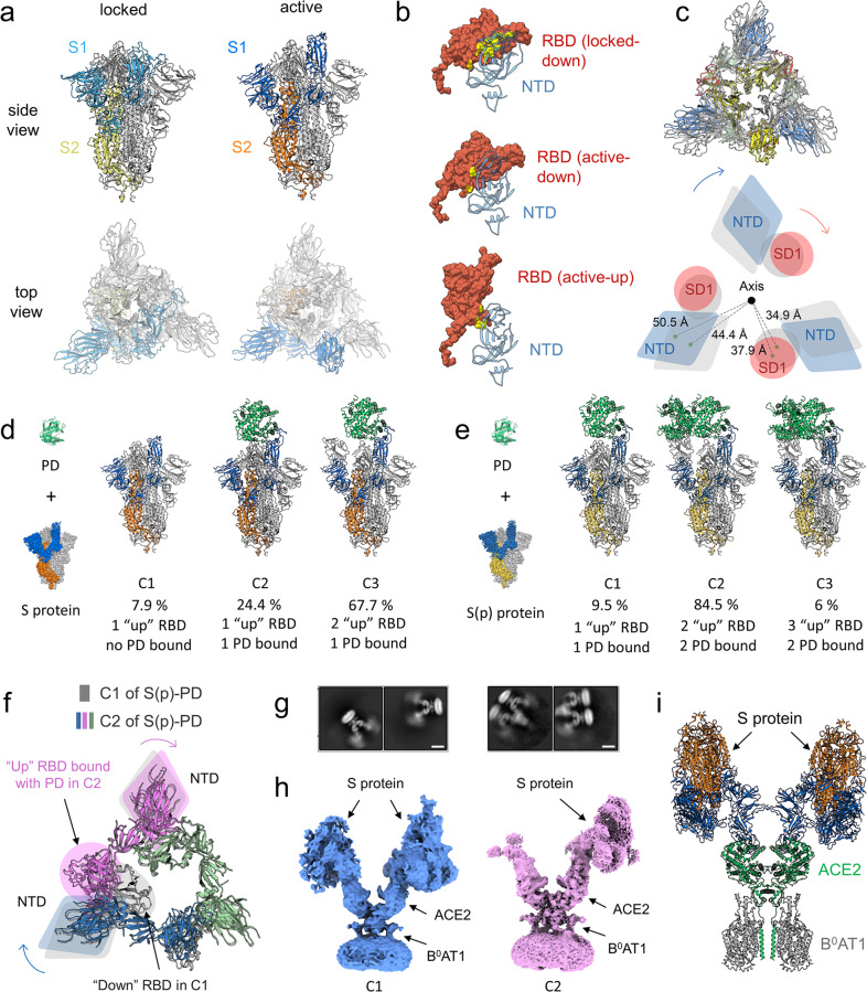
a Structures of the S protein. Left panel, locked conformation; right panel, active conformation with one RBD domain in the “up” position. b Decrease of contact area between RBD and NTD from the “down” RBD in the locked conformation (locked-down, upper panel), to the “down” RBD in the active conformation (active-down, middle panel), and to the “up” RBD in the active conformation (active-up, lower panel). c Structural comparison reveals a clockwise twist from the locked to the active conformation (upper panel), which loosens the contact between NTD and SD1 domain and increases the distance of these domains to the central axis. A schematic diagram is shown in the lower panel. Locked conformation is shown in gray. Active conformation is colored by domain, in which NTD is blue, RBD is yellow, SD1 is red and SD2 is green. d, e Different conformations of the S protein (d) and the S(p) protein (e) incubated with PD are shown, respectively. C1, C2 and C3 refer to conformation 1, 2 and 3, respectively. f Comparison between C1 and C2 of the S(p)–PD complex. Only RBD and NTD are shown for clarity. The binding of the second PD to RBD in C2 causes slight shift of NTDs both in the same protomer (violet) and in the anticlockwise protomer (blue). All protomers in C1 are colored in gray, whereas three protomers in C2 are colored in blue, violet and green, respectively. Circle and ellipse, RBD; diamond, NTD. g 2D class averages of the S–ACE2–B0AT1 ternary complex are shown. Scale bar, 10 nm. h Cryo-EM maps of conformation 1 (C1) and 2 (C2) of the S–ACE2–B0AT1 ternary complex. i Docking model of conformation 1 of the S–ACE2–B0AT1 ternary complex.
References
Publication types
MeSH terms
Substances
Grants and funding
LinkOut - more resources
Full Text Sources
Other Literature Sources
Molecular Biology Databases
Miscellaneous
