. 2021 Feb 8;10(3):e697-e703.
doi: 10.1016/j.eats.2020.10.059. eCollection 2021 Mar.

Segmental Meniscus Allograft Transplantation

Affiliations

Segmental Meniscus Allograft Transplantation

Max N Seiter et al. Arthrosc Tech. .

Abstract

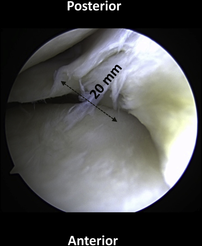
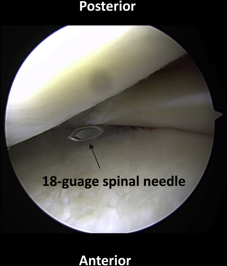
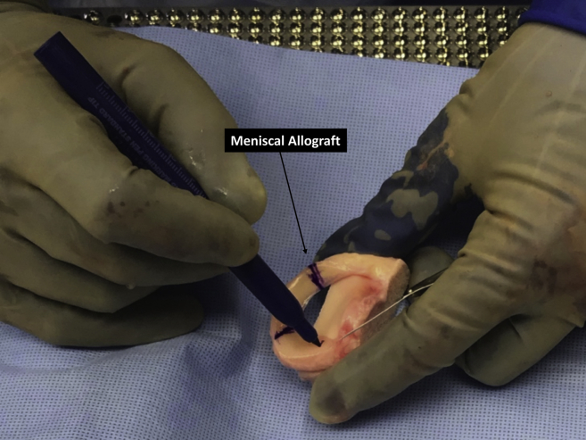
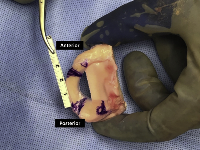
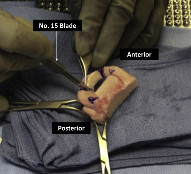
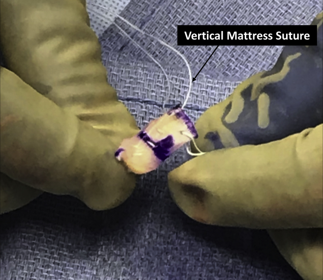
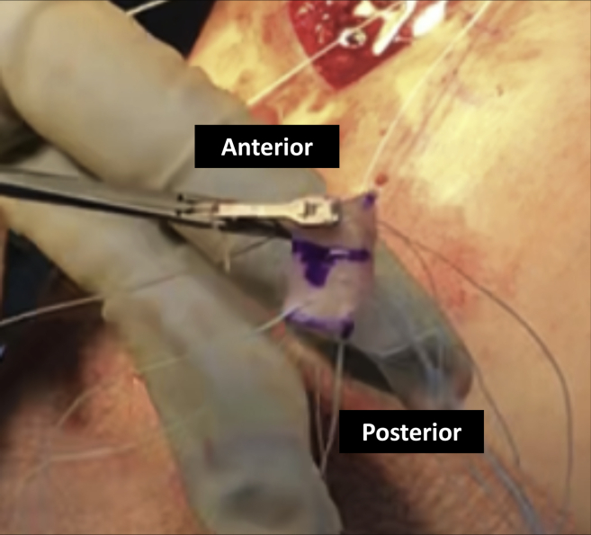
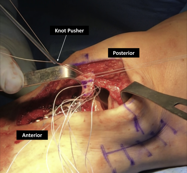
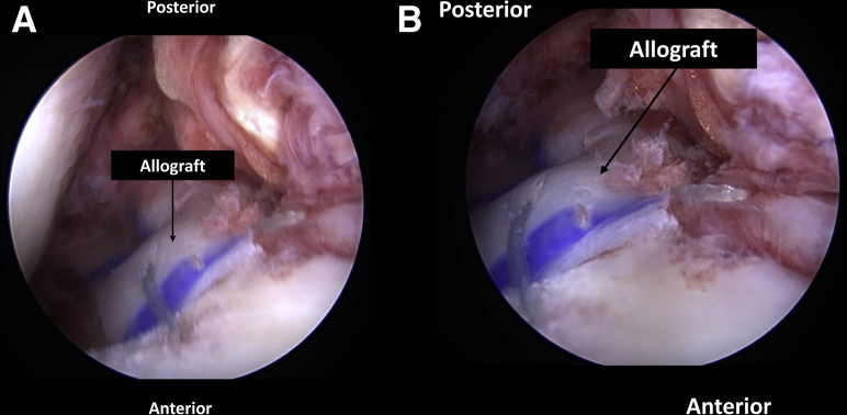
Meniscal tears treated with partial meniscectomies have been shown to significantly increase contract pressures within the tibiofemoral joint, and a complete focal meniscal deficiency may render the entirety of the meniscus functionally incompetent. Although various techniques of meniscal transplantation have been described, these techniques may require the excision of a considerable amount of healthy meniscal tissue. Furthermore, failures continue to frequently occur. Therefore, attempts to restoring normal knee kinematics and biomechanical forces are essential. Segmental meniscus allograft transplantations may offer the advantage of a robust repair by both maintaining knee biomechanics and biology while maximizing preservation of native meniscal tissue. Also, most meniscal deficiency involves only a portion of the meniscus, and thus we developed this technique to segmentally transplant only the deficient portion. The purpose of this Technical Note is to describe a technique of segmental medial meniscus allograft transplantation in a patient with focal medial meniscus deficiency.


Figures

Fig 1
Fig 1
Diagnostic arthroscopy viewing from the anterolateral portal shows a radial degenerative tear of the meniscus with healthy meniscus anterior and posterior to it. Rather than performing a near-total meniscectomy, a segmental portion can be removed and prepared for a segmental transplantation. The patient is positioned supine on the operating room table.
Fig 2
Fig 2
With the arthroscope in the anterolateral portal, a spinal needle is used for localization of the segmental transplant location. A small medial parapatellar arthrotomy is made.
Fig 3
Fig 3
After preparation of the recipient site is complete and the defect measured, the allograft meniscus is marked to the same size on the back table.
Fig 4
Fig 4
The allograft meniscus is measured along its periphery, verifying appropriate size for the segmental transplant. This measurement should equal the measured defect size at the periphery of the focal defect.
Fig 5
Fig 5
The allograft meniscus is cut to the appropriate size for the segmental transplantation. A No. 15 blade is used, while the assistant carefully stabilizes the graft.
Fig 6
Fig 6
While still on the back table, 5 vertical mattress sutures are then passed through the peripheral portion of the meniscus in preparation for the later outside-in repair over the capsule, as well as through the native meniscus. The authors find the use of an arthroscopic suture passer to be ideal for this task. In addition, 2 sutures were placed in the posterior aspect of the intact meniscus for repair.
Fig 7
Fig 7
The graft is then moved to the operating table. The sutures that were already passed through the native meniscus are then passed with an arthroscopic suture passer through the posterior portion of the segmental transplant.
Fig 8
Fig 8
An arthroscopic knot pusher is used to pass the meniscus through the medial arthrotomy and tensioned down, delivering the meniscus into the defect using a tension slide technique. The knee may be flexed as needed to facilitate delivery. An arthroscopic suture passer is then used to repair the meniscal graft to the capsule along the course of the arcuate ligaments. Additional inverted mattress sutures were then placed at the anterior rim of the meniscus. For further fixation, a 3-mm SutureTak knotless anchor (Arthrex, Naples, FL, U.S.A.) was placed anteriorly.
Fig 9
Fig 9
Final arthroscopic images viewed from the anterolateral portal after (A) suture fixation through the capsule, as done in an outside in meniscal repair. (B) Sutures are also seen tied to the adjacent anterior and posterior native meniscus

References

    1. Allaire R., Muriuki M., Gilbertson L., Harner C.D. Biomechanical consequences of a tear of the posterior root of the medial meniscus: Similar to total meniscectomy. J Bone Joint Surg Am. 2008;90:1922–1931. - PubMed
    1. Cox J.S., Nye C.E., Schaefer W.W., Woodstein I.J. The degenerative effects of partial and total resection of the medial meniscus in dogs' knees. Clin Orthop Relat Res. 1975;109:178–183. - PubMed
    1. Ichiba A., Kishimoto I. Effects of articular cartilage and meniscus injuries at the time of surgery on osteoarthritic changes after anterior cruciate ligament reconstruction in patients under 40 years old. Arch Orthop Trauma Surg. 2009;129:409–415. - PubMed
    1. Koh J.L., Yi S.J., Ren Y., Zimmerman T.A., Zhang L.Q. Tibiofemoral contact mechanics with horizontal cleavage tear and resection of the medial meniscus in the human knee. J Bone Joint Surg Am. 2016;98:1829–1836. - PubMed
    1. Lee B.S., Bin S.I., Kim J.M. Articular cartilage degenerates after subtotal/total lateral meniscectomy but radiographic arthrosis progression is reduced after meniscal transplantation. Am J Sports Med. 2016;44:159–165. - PubMed

LinkOut - more resources