Review
. 2021 Mar 2:9:645235.
doi: 10.3389/fcell.2021.645235. eCollection 2021.

Cellular and Supracellular Planar Polarity: A Multiscale Cue to Elongate the Drosophila Egg Chamber

Affiliations
Review

Cellular and Supracellular Planar Polarity: A Multiscale Cue to Elongate the Drosophila Egg Chamber

Anna Popkova et al. Front Cell Dev Biol. .

Abstract

Tissue elongation is known to be controlled by oriented cell division, elongation, migration and rearrangement. While these cellular processes have been extensively studied, new emerging supracellular mechanisms driving tissue extension have recently been unveiled. Tissue rotation and actomyosin contractions have been shown to be key processes driving Drosophila egg chamber elongation. First, egg chamber rotation facilitates the dorsal-ventral alignment of the extracellular matrix and of the cell basal actin fibers. Both fiber-like structures form supracellular networks constraining the egg growth in a polarized fashion thus working as 'molecular corsets'. Second, the supracellular actin fiber network, powered by myosin periodic oscillation, contracts anisotropically driving tissue extension along the egg anterior-posterior axis. During both processes, cellular and supracellular planar polarity provide a critical cue to control Drosophila egg chamber elongation. Here we review how different planar polarized networks are built, maintained and function at both cellular and supracellular levels in the Drosophila ovarian epithelium.

Keywords: actomyosin contractility; planar cell polarity; supracellular network; tissue elongation; tissue rotation.


Conflict of interest statement

The authors declare that the research was conducted in the absence of any commercial or financial relationships that could be construed as a potential conflict of interest.

Figures
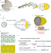
FIGURE 1
FIGURE 1
Egg chamber rotation and planar cell polarity during Drosophila oogenesis. (A) Schematic diagram illustrating Drosophila ovary, composed of 15–20 ovarioles. Each ovariole is composed of a string of egg chambers at different developmental stages. (B) Schematic diagram illustrating egg chamber morphology at stages 5–8. FC, follicle cells; NC, nurse cells; oocyte in yellow. Anterior-posterior (AP) and Dorsal-ventral (DV) axes are indicated. Green arrows mark the direction of tissue expansion, and blue arrows mark the corset-restrictive forces. The egg chambers expand their volume from early to late stages. With the corset-restrictive forces, the egg elongates along the AP-axis during their expansion processes. (C) Schematic representation of the cross-sections of egg chamber at stages 5–8. Arrows indicate the sense of tissue rotation. (D) Schematic representation of the molecular corsets at stage 5–8. The molecular corsets are composed of parallel fibrillary ECM at the BM and parallel actin bundles at the basal domain of follicular epithelial cells. (E) Schematic diagram showing the mechanisms driving tissue rotation and planar cell polarity during Drosophila oogenesis.
FIGURE 2
FIGURE 2
Oscillatory actomyosin contractions and planar cell polarity during Drosophila oogenesis. (A) Schematic diagram illustrating egg chamber morphology at stages 9–10. AP and DV axes are indicated. FC, follicle cells; NC, nurse cells; oocyte in yellow. The egg chamber elongates along the AP-axis. (B) Schematic representation of the cross-sections of the egg chamber at S9–S10. Arrows indicate forces directed along the AP axis. (C) Schematic representation of the actomyosin distribution at the basal side of follicle cells in contact during S9–S10, showing filopodia penetrating the actomyosin cortex of a neighboring cell and forming a supracellular contractile network. Arrows indicate contractile forces. (D) Schematic diagram showing the mechanisms driving actomyosin contractions and planar cell polarity during Drosophila oogenesis.

References

    1. Aigouy B., Farhadifar R., Staple D. B., Sagner A., Roper J. C., Julicher F., et al. (2010). Cell flow reorients the axis of planar polarity in the wing epithelium of Drosophila. Cell 142 773–786. 10.1016/j.cell.2010.07.042 - DOI - PubMed
    1. Baena-Lopez L. A., Baonza A., Garcia-Bellido A. (2005). The orientation of cell divisions determines the shape of Drosophila organs. Curr. Biol. 15 1640–1644. 10.1016/j.cub.2005.07.062 - DOI - PubMed
    1. Bailles A., Collinet C., Philippe J. M., Lenne P. F., Munro E., Lecuit T. (2019). Genetic induction and mechanochemical propagation of a morphogenetic wave. Nature 572 467–473. 10.1038/s41586-019-1492-9 - DOI - PMC - PubMed
    1. Banerjee D. S., Munjal A., Lecuit T., Rao M. (2017). Actomyosin pulsation and flows in an active elastomer with turnover and network remodeling. Nat. Commun. 8:1121. 10.1038/s41467-017-01130-1 - DOI - PMC - PubMed
    1. Barlan K., Cetera M., Horne-Badovinac S. (2017). Fat2 and lar define a basally localized planar signaling system controlling collective cell migration. Dev. Cell 40 467–477.e5. 10.1016/j.devcel.2017.02.003 - DOI - PMC - PubMed

LinkOut - more resources