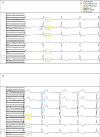
Progression of electrocardiogram changes in an untreated fabry disease: a case report
- PMID: 33738419
- PMCID: PMC7954241
- DOI: 10.1093/ehjcr/ytab045
Progression of electrocardiogram changes in an untreated fabry disease: a case report
Abstract
Background: Fabry disease (FD) is a rare lysosomal storage disorder with multiorgan manifestation and associated with an increased morbidity and mortality. Fabry cardiomyopathy includes left ventricular 'hypertrophy' (LVH), cardiac arrhythmias, and heart failure. We report a case of an untreated FD with characteristic findings in electrocardiogram (ECG) over a follow-up period of 10 years.
Case summary: A 53-year-old man with FD presented to our outpatient department. He suffered from symptomatic ventricular extrasystoles. Echocardiography detected LVH and reduced global longitudinal strain. Twelve years ago, first examination was conducted due to ventricular arrhythmias. Electrocardiogram showed a short PQ minus P-wave (PendQ) interval and negative T-waves. Over time, the number of leads with negative T-waves increased. Moreover, the echocardiography revealed a thickened left ventricular wall. Without any further examinations at that time, the patient was treated for arterial hypertension with proteinuria. Ten years after first symptoms appeared, FD was diagnosed utilizing cardiac magnetic resonance imaging and genetic tests. Hence, enzyme replacement therapy was initiated.
Discussion: The ECG is a fast diagnostic method and it may - even without additional organ manifestations - provide preliminary suspicion of FD. In particular, as shown in our case, a short PendQ and QT interval indicate FD. Over time, disease progression can be detected through ECG changes. T-waves correlate with an increasing LVH and a reduction in longitudinal function in echocardiographic examinations. Unexplained LVH must be followed by differential diagnosis. In case of confirmed FD, patients should be treated by multidisciplinary teams in experienced centres.
Keywords: Case report; Electrocardiogram; Enzyme replacement therapy; Fabry disease; Left ventricular hypertrophy.
© The Author(s) 2021. Published by Oxford University Press on behalf of the European Society of Cardiology.
Figures


Similar articles
-
Difficulties in Diagnosing Fabry Disease in Patients with Unexplained Left Ventricular Hypertrophy (LVH): Is the Novel GLA Gene Mutation a Pathogenic Mutation or Polymorphism?Balkan J Med Genet. 2023 Jul 31;26(1):43-50. doi: 10.2478/bjmg-2023-0010. eCollection 2023 Jul. Balkan J Med Genet. 2023. PMID: 37576794 Free PMC article.
-
Systolic and diastolic function assessment in fabry disease patients using speckle-tracking imaging and comparison with conventional echocardiographic measurements.J Am Soc Echocardiogr. 2013 Dec;26(12):1407-14. doi: 10.1016/j.echo.2013.09.005. Epub 2013 Oct 11. J Am Soc Echocardiogr. 2013. PMID: 24125876
-
Value of electrocardiogram in the differentiation of hypertensive heart disease, hypertrophic cardiomyopathy, aortic stenosis, amyloidosis, and Fabry disease.Am J Cardiol. 2012 Feb 15;109(4):587-93. doi: 10.1016/j.amjcard.2011.09.052. Epub 2011 Nov 19. Am J Cardiol. 2012. PMID: 22105784
-
Fabry disease and its cardiac involvement.J Gen Fam Med. 2017 May 8;18(5):225-229. doi: 10.1002/jgf2.76. eCollection 2017 Oct. J Gen Fam Med. 2017. PMID: 29264031 Free PMC article. Review.
-
Fabry Disease: More than a Phenocopy of Hypertrophic Cardiomyopathy.J Clin Med. 2023 Nov 13;12(22):7061. doi: 10.3390/jcm12227061. J Clin Med. 2023. PMID: 38002674 Free PMC article. Review.
References
-
- Chan B, Adam DN. A review of Fabry disease. Skin Ther Lett 2018;23:4–6. - PubMed
-
- Seydelmann N, Wanner C, Störk S, Ertl G, Weidemann F. Fabry disease and the heart. Best Pract Res Clin Endocrinol Metab 2015;29:195–204. - PubMed
-
- Takenaka T, Teraguchi H, Yoshida A, Taguchi S, Ninomiya K, Umekita Y. et al. Terminal stage cardiac findings in patients with cardiac Fabry disease: an electrocardiographic, echocardiographic, and autopsy study. J Cardiol 2008;51:50–59. - PubMed
-
- Kouris NT, Kontogianni DD, Pavlou MT, Babalis DK. Atrioventricular conduction disturbances in a young patient with Fabry's disease without other signs of cardiac involvement. Int J Cardiol 2005;99:327–328. - PubMed
Publication types
LinkOut - more resources
Full Text Sources
Other Literature Sources
