

Submitral aneurysm of varied aetiologies: a case series
- PMID: 33738423
- PMCID: PMC7954274
- DOI: 10.1093/ehjcr/ytab066
Submitral aneurysm of varied aetiologies: a case series
Abstract
Background: Submitral aneurysm is a rare disease initially described in the African population. It is usually considered congenital in origin, due to a defect in the posterior portion of the mitral annulus. However, it can be seen in other diseases like ischaemic heart disease, rheumatic heart disease, infective endocarditis, tuberculosis, and syphilis.
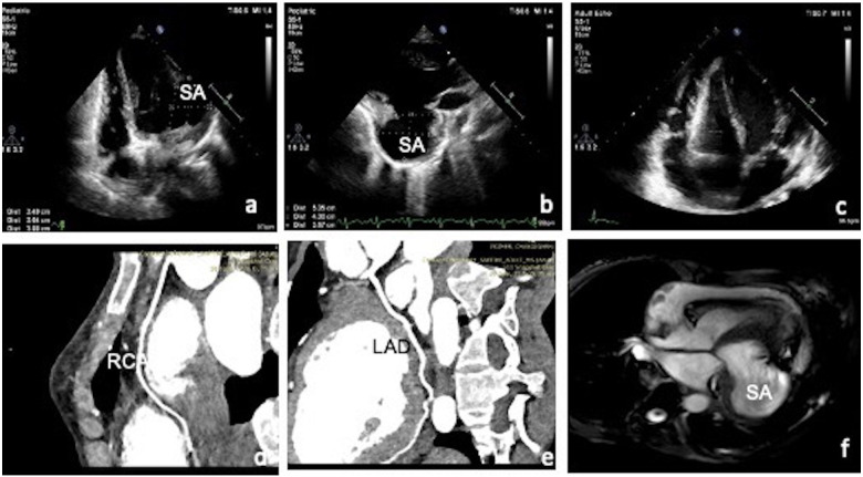
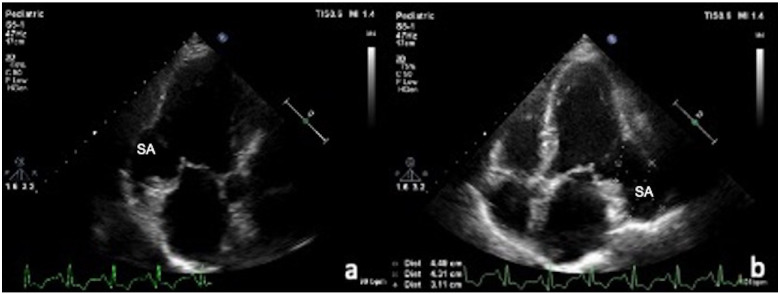
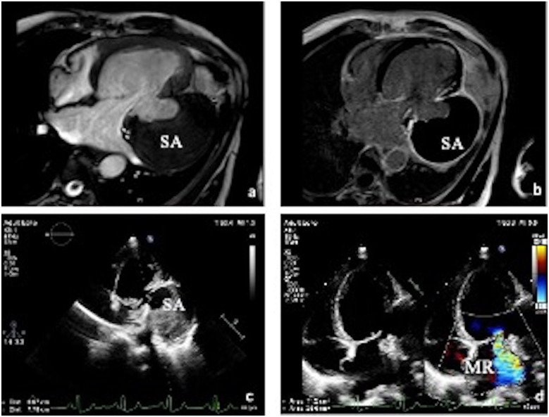
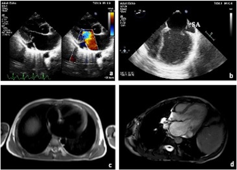
Case presentation: Case 1 was a 29-year-old female, hypertensive undergoing maintenance haemodialysis for chronic kidney disease and on anti-tubercular therapy. She was found to have a large submitral aneurysm with severe mitral regurgitation, moderate left ventricular dysfunction, and pericardial effusion on echocardiogram. Case 2 was a 58-year-old gentleman presented with inferior wall ST-elevation myocardial infarction and was thrombolyzed with streptokinase for the same. Echocardiogram done 6 months later for evaluation of dyspnoea showed a large inferobasal aneurysm. Case 3 was a 56-year-old hypertensive presented with dyspnoea on exertion and echocardiogram showed a large posterolateral region with transmural late gadolinium enhancement. Case 4 was a 13-year-old boy presented with fever and cerebrovascular accident. Echocardiogram revealed vegetation in the mitral valve and a small submitral aneurysm with vegetation inside it.
Discussion: Submitral aneurysm is usually considered congenital in origin. However, it can be due to ischaemic heart disease, rheumatic heart disease, Takayasu arteritis, and tuberculosis. Top dimensional echocardiogram is the investigation of choice. Cardiac magentic resonance imaging helps in identifying the underlying aetiology and delineating the surrounding structures.
Keywords: Case series; Coronary artery disease; Infective endocarditis; Submitral aneurysms; Tuberculosis.
© The Author(s) 2021. Published by Oxford University Press on behalf of the European Society of Cardiology.
Figures

References
-
- Abrahams DG, Barton CJ, Cockshott WP, Edington GM, Weaver EJ.. Annular subvalvular left ventricular aneurysms. Q J Med 1962;31:345–360. - PubMed
-
- Rose AG, Folb J, Sinclair-Smith CC, Schneider JW.. Idiopathic annular submitral aneurysm associated with Takayasu's aortitis. A report of two cases. Arch Pathol Lab Med 1995;119:831–835. - PubMed
-
- Kanabar K, Prasad K, Rani P, Kaur N, Santosh K, Mehrotra S.. Large infero-basal left ventricular aneurysm with organized thrombus. J Echocardiogr 2020;18:262–264. - PubMed
-
- Cheng TO. Ventricular aneurysm and coronary artery disease: the hen and the egg? Cathet Cardiovasc Diagn 1997;41:468–468. - PubMed
Publication types
LinkOut - more resources
Full Text Sources
Other Literature Sources