

Macular pigment optical density in central serous chorioretinopathy
- PMID: 33738428
- PMCID: PMC7934065
- DOI: 10.1177/2515841421997195
Macular pigment optical density in central serous chorioretinopathy
Abstract
Purpose: The aim of our study was to evaluate the macular pigment optical density in patients with acute and chronic central serous chorioretinopathy and to describe the association between central retinal thickness and choroidal thickness with the macular pigment optical density.
Materials and methods: Eyes with acute central serous chorioretinopathy and chronic central serous chorioretinopathy (patients, who were diagnosed as having disease activity for 6 months) were included in this study. Macular pigment was measured using the heterochromatic flicker technique of the MPS II device for both eyes in patients with acute and chronic central serous chorioretinopathy and in control subjects.
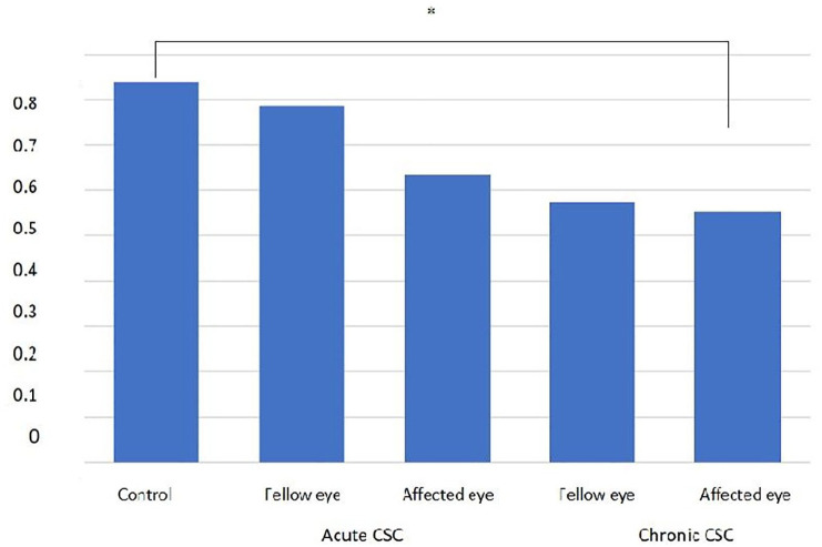
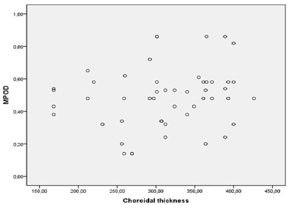
Results: Twenty-seven eyes with acute central serous chorioretinopathy, 23 eyes with chronic central serous chorioretinopathy, and 25 control eyes were enrolled. The mean macular pigment optical density in chronic central serous chorioretinopathy (0.480 ± 0.16 density unit (95% confidence interval: 0.390-0.570) was found to be significantly lower than in the control eyes (0.571 ± 0.128 density unit) (95% confidence interval: 0.480-0.670) (p = 0.007). In correlation analysis, no significant association was detected between the central retinal thickness, choroidal thickness, and macular pigment optical density values in central serous chorioretinopathy group (p = 0.31, p = 0.71).
Conclusion: Macular pigment optical density levels were significantly lower in chronic central serous chorioretinopathy patients than in controls, possibly due to degeneration of the neurosensorial retina, as a result of the long-term persistence of subretinal fluid. There was not a significant correlation between choroidal thickness and macular pigment optical density levels in central serous chorioretinopathy group.
Keywords: Central retinal thickness; central serous chorioretinopathy; choroidal thickness; macular pigment optical density.
© The Author(s), 2021.
Conflict of interest statement
Conflict of interest statement: The authors declared no potential conflicts of interest with respect to the research, authorship, and/or publication of this article.
Figures


References
-
- Yannuzzi LA. Central serous chorioretinopathy: a personal perspective. Am J Ophthalmol 2010; 149: 361–363.e1. - PubMed
-
- Haimovici R, Koh S, Gagnon DR, et al. Risk factors for central serous chorioretinopathy: a case–control study. Ophthalmology 2004; 111: 244–249. - PubMed
-
- Ruiz-Medrano J, Pellegrini M, Cereda MG, et al. Choroidal characteristics of acute and chronic central serous chorioretinopathy using enhanced depth imaging optical coherence tomography. Eur J Ophthalmol 2017; 27: 476–480. - PubMed
-
- Imamura Y, Fujiwara T, Margolis R, et al. Enhanced depth imaging optical coherence tomography of the choroid in central serous chorioretinopathy. Retina 2009; 29: 1469–1473. - PubMed
LinkOut - more resources
Full Text Sources
Other Literature Sources
