Review
. 2022 May;110(5):592-604.
doi: 10.1007/s00223-021-00836-1. Epub 2021 Mar 18.

Disuse Osteoporosis: Clinical and Mechanistic Insights

Affiliations
Review

Disuse Osteoporosis: Clinical and Mechanistic Insights

Tim Rolvien et al. Calcif Tissue Int. 2022 May.

Abstract

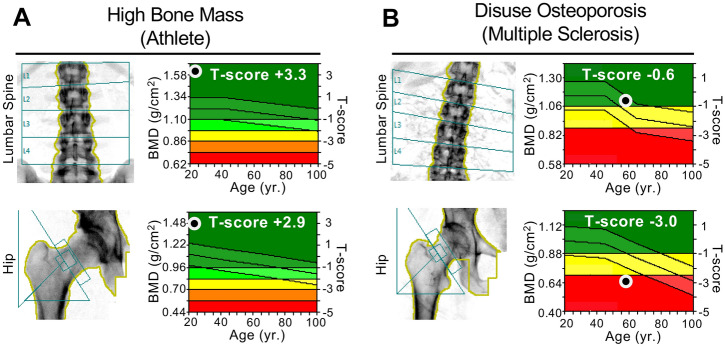
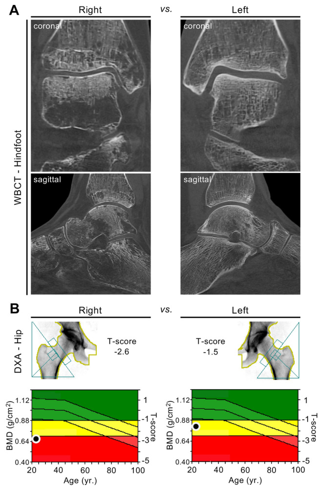
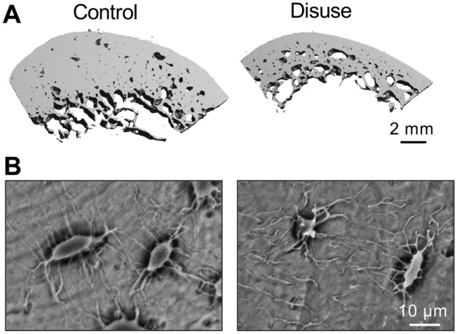
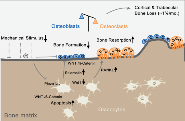
Disuse osteoporosis describes a state of bone loss due to local skeletal unloading or systemic immobilization. This review will discuss advances in the field that have shed light on clinical observations, mechanistic insights and options for the treatment of disuse osteoporosis. Clinical settings of disuse osteoporosis include spinal cord injury, other neurological and neuromuscular disorders, immobilization after fractures and bed rest (real or modeled). Furthermore, spaceflight-induced bone loss represents a well-known adaptive process to microgravity. Clinical studies have outlined that immobilization leads to immediate bone loss in both the trabecular and cortical compartments accompanied by relatively increased bone resorption and decreased bone formation. The fact that the low bone formation state has been linked to high levels of the osteocyte-secreted protein sclerostin is one of the many findings that has brought matrix-embedded, mechanosensitive osteocytes into focus in the search for mechanistic principles. Previous basic research has primarily involved rodent models based on tail suspension, spaceflight and other immobilization methods, which have underlined the importance of osteocytes in the pathogenesis of disuse osteoporosis. Furthermore, molecular-based in vitro and in vivo approaches have revealed that osteocytes sense mechanical loading through mechanosensors that translate extracellular mechanical signals to intracellular biochemical signals and regulate gene expression. Osteocytic mechanosensors include the osteocyte cytoskeleton and dendritic processes within the lacuno-canalicular system (LCS), ion channels (e.g., Piezo1), extracellular matrix, primary cilia, focal adhesions (integrin-based) and hemichannels and gap junctions (connexin-based). Overall, disuse represents one of the major factors contributing to immediate bone loss and osteoporosis, and alterations in osteocytic pathways appear crucial to the bone loss associated with unloading.

Keywords: Bone loss; Immobilization; Microstructure; Osteocyte; Unloading.


Conflict of interest statement

Tim Rolvien and Michael Amling declare that they have no conflict of interest.

Figures

Fig. 1
Fig. 1
Clinical examples of two patients with high vs. low BMD in response to intense physical exercise vs. unloading. a DXA scans of a professional athlete (soccer player) with high BMD values in both the lumbar spine and hip. b DXA scans of an immobilized patient with multiple sclerosis showing low BMD values, which were more severe in the hip than the lumbar spine, meeting the World Health Organization’s (WHO) definition of osteoporosis in the hip (i.e., T-score ≤ − 2.5)
Fig. 2
Fig. 2
Local deterioration of bone mass after fracture and subsequent unilateral disuse of the right limb. a Cone-beam computed tomography (CBCT) images of the right foot compared to the left foot (coronal and sagittal reconstruction) in a 22-year-old female patient obtained eight months after suffering from an ankle fracture with subsequent unilateral unloading of the right limb. Focal osteolytic changes (often referred to as “local disuse osteopenia/osteoporosis”) are visible. b DXA scans demonstrating the differences in BMD between the right and left proximal femur (T-score − 2.6 vs. − 1.5). Bilateral HR-pQCT scans of the distal tibiae were also performed and showed cortical and trabecular bone loss syndrome on the unloaded side compared to the contralateral (loaded) side (cortical thickness − 28%, bone volume per tissue volume − 7%)
Fig. 3
Fig. 3
Micromorphological changes in cortical bone after immobilization. a µ-CT images demonstrating profound cortical changes, including a decrease in cortical thickness and an increase in cortical porosity, in individuals after long-term bed rest. b SEM images of acid-etched plastic-embedded cortical bone sections revealing decreased canalicular connectivity of osteocytes after immobilization, underlining the role of osteocytes in mechanotransduction. Images were obtained from representative samples obtained in the context of a previous study [79]
Fig. 4
Fig. 4
Schematic model of the molecular pathways involved in skeletal unloading. Mechanosensation in osteocytes is mediated by the lacuno-canalicular system (LCS) and via ion channels of the Piezo family (primarily Piezo1), among others. Inactivation of Piezo1 leads to decreased Wnt1 expression. Furthermore, sclerostin secretion by osteocytes is increased in response to unloading, which inhibits Wnt/β-catenin and results in suppressed osteoblast activity. Unloading also leads to increased osteocyte apoptosis (partly via Wnt/β-catenin signaling) and increased RANKL expression, promoting increased bone resorption

References

    1. Zaidi M. Skeletal remodeling in health and disease. Nat Med. 2007;13:791–801. doi: 10.1038/nm1593. - DOI - PubMed
    1. Wolff J. Das Gesetz der Transformation der Knochen. Berlin: Hirschwald; 1892.
    1. Huddleston AL, Rockwell D, Kulund DN, Harrison RB. Bone mass in lifetime tennis athletes. JAMA. 1980;244:1107–1109. doi: 10.1001/jama.1980.03310100025022. - DOI - PubMed
    1. Schaffler MB, Cheung WY, Majeska R, Kennedy O. Osteocytes: master orchestrators of bone. Calcif Tissue Int. 2014;94:5–24. doi: 10.1007/s00223-013-9790-y. - DOI - PMC - PubMed
    1. Poole KE, van Bezooijen RL, Loveridge N, Hamersma H, Papapoulos SE, Lowik CW, Reeve J. Sclerostin is a delayed secreted product of osteocytes that inhibits bone formation. FASEB J. 2005;19:1842–1844. doi: 10.1096/fj.05-4221fje. - DOI - PubMed

Publication types