Review
. 2021 Mar 18;21(3):20.
doi: 10.1007/s11882-020-00981-x.

A TRiP Through the Roles of Transient Receptor Potential Cation Channels in Type 2 Upper Airway Inflammation

Affiliations
Review

A TRiP Through the Roles of Transient Receptor Potential Cation Channels in Type 2 Upper Airway Inflammation

Wout Backaert et al. Curr Allergy Asthma Rep. .

Abstract

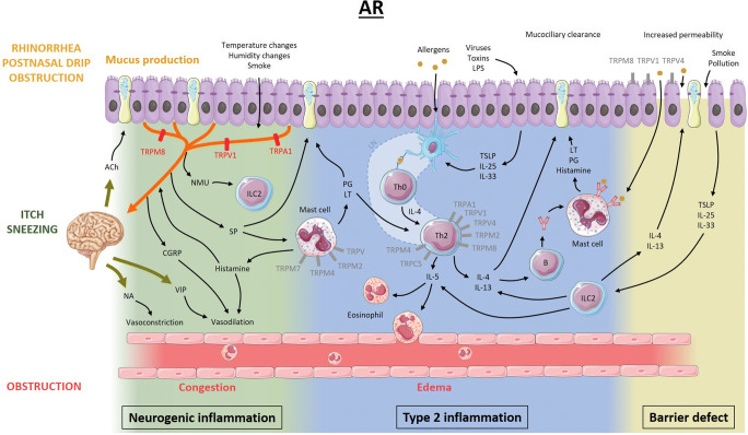
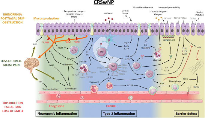
Purpose of review: Despite their high prevalence, the pathophysiology of allergic rhinitis (AR) and chronic rhinosinusitis (CRS) remains unclear. Recently, transient receptor potential (TRP) cation channels emerged as important players in type 2 upper airway inflammatory disorders. In this review, we aim to discuss known and yet to be explored roles of TRP channels in the pathophysiology of AR and CRS with nasal polyps.

Recent findings: TRP channels participate in a plethora of cellular functions and are expressed on T cells, mast cells, respiratory epithelial cells, and sensory neurons of the upper airways. In chronic upper airway inflammation, TRP vanilloid 1 is mostly studied in relation to nasal hyperreactivity. Several other TRP channels such as TRP vanilloid 4, TRP ankyrin 1, TRP melastatin channels, and TRP canonical channels also have important functions, rendering them potential targets for therapy. The role of TRP channels in type 2 inflammatory upper airway diseases is steadily being uncovered and increasingly recognized. Modulation of TRP channels may offer therapeutic perspectives.

Keywords: Allergic rhinitis; Chronic rhinosinusitis; Mast cell; Nasal hyperreactivity; Respiratory epithelial cell; T cell; Transient receptor potential; Type 2 inflammation.


Conflict of interest statement

The authors declare no conflicts of interest relevant to this manuscript.

Figures

Fig. 1
Fig. 1
Proposed model of the pathophysiology of allergic rhinitis and the potential role of TRP channels. Allergens presented by dendritic cells induce maturation of Th0 cells to Th2 cells in lymph nodes. Pro-inflammatory mediators such as IL-4, IL-5, and IL-13 are released by Th2 cells and activated type 2 innate lymphoid cells and induce eosinophil recruitment/activation and production of monoclonal allergen-specific IgE. In sensitized individuals, mast cell mediators are released upon binding of allergens to allergen-specific IgE and induce mucus production, vasodilation, and plasma extravasation leading to nasal congestion and edema, ultimately resulting in nasal obstruction, rhinorrhea, and post-nasal drip. On the other hand, epithelial mediators can activate dendritic cells and type 2 innate lymphoid cells. Activation of sensory afferent neurons by histamine results in nasal itch and sneezing. Upon neuronal activation by endogenous or exogenous triggers, the signal travels to the central nervous system, inducing parasympathetic (increased mucus production and vasodilation) or orthosympathetic (vasoconstriction) responses (orthodromic pathway). However, in case of nasal hyperreactivity, neuropeptides are released directly from afferent nerves (antidromic pathway). Lastly, not only environmental factors, but also inflammatory mediators disrupt the epithelial barrier function, increasing epithelial permeability for potentially noxious stimuli. The role of TRPV1, TRPA1, and TRPM8 in neurogenic inflammation has been studied the most. However, TRP channels present on other cells could be potential therapeutic targets as well (gray). Ach, acetylcholine; NA, noradrenaline; VIP, vasoactive intestinal peptide; CGRP, calcitonin gene-related peptide; NMU, neuromedin U; SP, substance P; ILC2, type 2 innate lymphoid cell; PG, prostaglandins; LT, leukotrienes; LN, lymph node; IL, interleukin; LPS, lipopolysaccharide; TSLP, thymic stromal lymphopoietin
Fig. 2
Fig. 2
Proposed model of the pathophysiology of chronic rhinosinusitis with nasal polyps and the potential role of TRP channels. Antigens presented by dendritic cells initiate development of Th2 cells that release IL-4, IL-5, and IL-13. IL-5 recruits and activates eosinophils, which ultimately leads to fibrin cross-linking and polyp formation. IL-4 and IL-13 stimulate B cells to produce not only monoclonal IgE to Staphylococcus aureus antigens but also polyclonal autoantibodies leading to complement activation at the basement membrane of the epithelium. Together with protease activity from allergens, environmental factors, and inflammatory mediators, this leads to disruption of the epithelial barrier, facilitating penetration of environmental stimuli. Activated epithelial cells release pro-inflammatory mediators, which can activate dendritic cells and type 2 innate lymphoid cells. Much like in AR, the nervous system participates in the inflammatory process through neuro-immune interactions, modulating the immunological battlefield. Type 2 inflammatory mediators can activate and potentially sensitize sensory afferent neurons, while neuropeptides can activate mast cells and type 2 innate lymphoid cells (ILC2). Moreover, central reflexes regulate vascular tone and mucosal gland activity. The role of TRPV1, TRPA1, and TRPM8 in neurogenic inflammation has been studied the most. However, TRP channels present on other cells could be potential therapeutic targets as well (gray). Ach, acetylcholine; NA, noradrenaline; VIP, vasoactive intestinal peptide; CGRP, calcitonin gene-related peptide; NMU, neuromedin U; SP, substance P; ILC2, type 2 innate lymphoid cell; PG, prostaglandins; LT, leukotrienes; LN, lymph node; IL, interleukin; LPS, lipopolysaccharide; TSLP, thymic stromal lymphopoietin; CCL23, chemokine (C-C motif) ligand 23; FXIIIa, activated coagulation factor 13; t-PA, tissue plasminogen activator

Similar articles

Cited by

References

    1. • De Greve G, Hellings PW, Fokkens WJ, Pugin B, Steelant B, Seys SF. Endotype-driven treatment in chronic upper airway diseases. Clin Transl Allergy. 2017;7(22). Concise review covering the importance of endotyping and phenotyping in rhinitis and rhinosinusitis. - PMC - PubMed
    1. Hellings PW, Klimek L, Cingi C, Agache I, Akdis C, Bachert C, Bousquet J, Demoly P, Gevaert P, Hox V, Hupin C, Kalogjera L, Manole F, Mösges R, Mullol J, Muluk NB, Muraro A, Papadopoulos N, Pawankar R, Rondon C, Rundenko M, Seys SF, Toskala E, van Gerven L, Zhang L, Zhang N, Fokkens WJ. Non-allergic rhinitis: position paper of the European Academy of Allergy and Clinical Immunology. Allergy. 2017;72(11):1657–1665. - PubMed
    1. •• Fokkens WJ, Lund VJ, Hopkins C, Hellings PW, Kern R, Reitsma S, et al. European position paper on rhinosinusitis and nasal polyps 2020. Rhinology. 2020;58(S29):1–464. Hallmark paper on the pathophysiology and diagnostic and therapeutic approaches towards rhinosinusitis. - PubMed
    1. Sin B, Togias A. Pathophysiology of allergic and nonallergic rhinitis. Proc Am Thorac Soc. 2011;8:106–114. - PubMed
    1. Sarin S, Undem B, Sanico A, Togias A. The role of the nervous system in rhinitis. J Allergy Clin Immunol. 2006;118(5):999–1014. - PubMed

Publication types

Substances