Comparative Study
. 2021 Jun 1;42(8):2332-2346.
doi: 10.1002/hbm.25368. Epub 2021 Mar 19.

Brain age prediction: A comparison between machine learning models using region- and voxel-based morphometric data

Affiliations
Comparative Study

Brain age prediction: A comparison between machine learning models using region- and voxel-based morphometric data

Lea Baecker et al. Hum Brain Mapp. .

Abstract

Brain morphology varies across the ageing trajectory and the prediction of a person's age using brain features can aid the detection of abnormalities in the ageing process. Existing studies on such "brain age prediction" vary widely in terms of their methods and type of data, so at present the most accurate and generalisable methodological approach is unclear. Therefore, we used the UK Biobank data set (N = 10,824, age range 47-73) to compare the performance of the machine learning models support vector regression, relevance vector regression and Gaussian process regression on whole-brain region-based or voxel-based structural magnetic resonance imaging data with or without dimensionality reduction through principal component analysis. Performance was assessed in the validation set through cross-validation as well as an independent test set. The models achieved mean absolute errors between 3.7 and 4.7 years, with those trained on voxel-level data with principal component analysis performing best. Overall, we observed little difference in performance between models trained on the same data type, indicating that the type of input data had greater impact on performance than model choice. All code is provided online in the hope that this will aid future research.

Keywords: biological ageing; healthy ageing; machine learning; regression analysis; support vector machine.


Conflict of interest statement

The authors declare no conflict of interest.

Figures
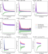
FIGURE 1
FIGURE 1
MAE of region‐ and voxel‐based SVR, RVR, and GPR models with or without PCA for the training set size compared to chance level (7.5 years; black dotted line). MAE is shown for the performance within the training (red line) and test set (green line) of the CV (Site 1) and in the independent test set (Site 2; blue line). The confidence intervals (shaded areas) for the different size of the data sets were calculated using bootstrap analysis. Note that bootstrap training samples were selected to be age‐ and sex‐homogeneous of increasing size with the minimum of one man and one woman per age and maximum of 20 men and 20 women per age. For the voxel‐based models with PCA, data sets with <150 subjects could not be assessed, because the PCA algorithm requires more samples than principal components. Furthermore, training set sizes above 500 were not calculated due limited time and computational resources
FIGURE 2
FIGURE 2
A decision tree for researchers choosing the most suitable brain age prediction model for their project. The ranking is inferred from our experience developing the models as well as the results of our investigation. These recommendations are thus built on the UK Biobank data set and our specific computational resources, so any application to other projects should be done with caution. The models in this study were developed using a high‐end consumer‐grade desktop computer with a 16‐core (32‐processes) CPU @ 3.40 GHz utilising 128 GB RAM. The voxel‐based models with PCA took 1–2 weeks to train, while the voxel‐based models without PCA took <1 day. The region‐based models took <1 hr to train

References

    1. Alexander, D. L. J. , Tropsha, A. , & Winkler, D. A. (2015). Beware of R2: Simple, unambiguous assessment of the prediction accuracy of QSAR and QSPR models. Journal of Chemical Information and Modeling, 55, 1316–1322. 10.1021/acs.jcim.5b00206 - DOI - PMC - PubMed
    1. Ashburner, J. (2007). A fast diffeomorphic image registration algorithm. NeuroImage, 38, 95–113. 10.1016/j.neuroimage.2007.07.007 - DOI - PubMed
    1. Avants, B. B. , Epstein, C. L. , Grossman, M. , & Gee, J. C. (2008). Symmetric diffeomorphic image registration with cross‐correlation: Evaluating automated labeling of elderly and neurodegenerative brain. Medical Image Analysis, 12, 26–41. 10.1016/j.media.2007.06.004 - DOI - PMC - PubMed
    1. Avants, B. B. , Tustison, N. , & Song, G. (2009). Advanced normalization tools (ANTS). Insight Journal, 1–35. Retrieved from ftp://ftp3.ie.freebsd.org/pub/sourceforge/a/project/ad/advants/Documenta...
    1. Avants, B. B. , Tustison, N. J. , Song, G. , Cook, P. A. , Klein, A. , & Gee, J. C. (2011). A reproducible evaluation of ANTs similarity metric performance in brain image registration. NeuroImage, 54(3), 2033–2044. 10.1016/j.neuroimage.2010.09.025 - DOI - PMC - PubMed

Publication types