. 2021 Apr;102(4):001580.
doi: 10.1099/jgv.0.001580.

Application of error-prone PCR to functionally probe the morbillivirus Haemagglutinin protein

Affiliations

Application of error-prone PCR to functionally probe the morbillivirus Haemagglutinin protein

Giulia Gallo et al. J Gen Virol. 2021 Apr.

Abstract

The enveloped morbilliviruses utilise conserved proteinaceous receptors to enter host cells: SLAMF1 or Nectin-4. Receptor binding is initiated by the viral attachment protein Haemagglutinin (H), with the viral Fusion protein (F) driving membrane fusion. Crystal structures of the prototypic morbillivirus measles virus H with either SLAMF1 or Nectin-4 are available and have served as the basis for improved understanding of this interaction. However, whether these interactions remain conserved throughout the morbillivirus genus requires further characterisation. Using a random mutagenesis approach, based on error-prone PCR, we targeted the putative receptor binding site for SLAMF1 interaction on peste des petits ruminants virus (PPRV) H, identifying mutations that inhibited virus-induced cell-cell fusion. These data, combined with structural modelling of the PPRV H and ovine SLAMF1 interaction, indicate this region is functionally conserved across all morbilliviruses. Error-prone PCR provides a powerful tool for functionally characterising functional domains within viral proteins.

Keywords: directed evolution; epPCR; morbillivirus; peste des petits ruminants virus; viral entry; viral evolution.


Conflict of interest statement

The authors declare that there are no conflicts of interest.

Figures
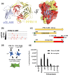
Fig. 1.
Fig. 1.
Error-prone PCR mutagenesis of PPRV H. (a) A model of ovine SLAM (oSLAM, blue ribbons) in complex with PPRV H (yellow and red ribbons), generated using the structure of MeV H in complex with marmoset SLAM (PDB ID 3ALX [9]) is shown in two orthogonal views. In the right-hand view the molecular surface of PPRV H is shown. Residues that were mutagenized by epPCR are coloured red. Model available at https://doi.org/10.17863/CAM.58532. (b) A simplified schematic of epPCR illustrating the correlation between increasing cycle number and the number of mutations (red crosses). (c) epPCR libraries of the PPRV H ORF were generated using Taq polymerase (Promega), as described in [13] with a 3′ primer including a NotI restriction site, and 64 cycles yielded 16 PCR products in total (represented by red bar; see Fig. S1a). These were subsequently pooled into four libraries (1–4, 5–8, 9–12 and 13–16; see Fig. S1b). Seperately, a faithful PCR copy of the N terminus (yellow) was amplified by standard high-fidelity PCR (using Kod polymerase, Merck) with a 5′ primer containing a T7 promoter sequence (green) and the NheI restriction site (blue). Lastly, overlap PCR was performed using the two external primers and Kod high-fidelity PCR to generate four libraries containing PCR products with full-length PPRV ORFs (yellow/red, L1-4, L5-8, L9-12 and L13-16 see Fig. S1b). (d) The quantifiable cell-cell fusion for PPRV relies on co-transfection of effector cells [E] with constructs expressing PPRV F and H proteins as well as half of a rLuc-GFP dual reporter [16]. These are co-cultured with target cells [T] expressing ovine SLAM and the corresponding half of the reporter. A cognate combination of FH and receptor leads to cell-cell fusion, reconstitution of the reporter and activation of the luciferase and GFP proteins. (e) Increasing epPCR mutagenesis of the PPRV H protein leads to reduced cell-cell fusion. Effector cells were transfected with the split reporter and no glycoprotein (No GP), plasmids bearing WT PPRV F and H (Plasmid), a plasmid expressing F and a PCR product expressing the PPRV H ORF (WT) amplified using the same external primers used in (c), or, the overlapped PCR products from this same epPCR protocol. After 2 days these were co-cultured with target cells expressing the corresponding half of the split reporter and either mock vector (No receptor) or a plasmid expressing ovine SLAMF1 (Ovine SLAM). Luciferase activity was measured using a cell permeable Renilla luciferase substrate (Coelenterazine) the following day. Assays were performed in biological triplicate with the mean and standard deviation plotted.
Fig. 2.
Fig. 2.
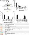
Screening of PPRV H epPCR libraries to identify individual mutants and link phenotype to genotype. (a) The pool selection and deconvolution strategy for identifying individual H mutants with modified cell-cell fusion activity (see main text for detailed explanation). (b) Screening of the pools L1-4A to L13-16-C (each containing 16 colonies) using a PPRV cell-cell fusion assay, as described in Fig. 1, identified a correlation between increased epPCR cycle number and decreased cell-cell fusion activity (similar to Fig. 1e). For WT a selection of colonies expressing the unmutated PPRV H was used (n=8 or as shown). (c) Pools L5-8A, L5-8C and L13-16B were taken forward for further analysis by sub-dividing the pools into four colonies per pool (e.g. L5-8A/1 to L5-8A/4) and re-examining their cell-cell fusion phenotype. (d, e) Based on these results pools L5-8A/1, L5-8C/2, L5-8C/3 and L13-16B/4 were separated into their individual colonies, re-screened in the cell-cell fusion assay and each mutant’s PPRV H ORF sequenced between nt 1335 and 1808 – the region targeted by epPCR – with any mutations to the PPRV H amino acid sequence calculated. Clone numbers in (e) are coloured based on mutation type and relative activity in the cell-cell assay (green=high activity relative to WT, orange=frameshift or premature stop codon, red=little or no activity relative to WT). Luciferase activity was measured using a cell permeable Renilla luciferase substrate (Coelenterazine). Assays were performed in biological triplicate with the mean and standard deviation plotted. Experiments in panel D were performed three separate times with a representative data set shown.
Fig. 3.
Fig. 3.
Examining the predicted structural configuration, electrophoretic mobility and glycosylation pattern of individual PPRV H mutants. (a, b) A model of ovine SLAM (oSLAM, blue ribbons) in complex with PPRV H (yellow ribbons) is shown in three orthogonal views. (a) Residues that could be mutated without abolishing PPRV H binding to oSLAM are shown in spacefill, coloured light green (L5-8C/3D) or dark green (L5-8C/2D). (b) Residues that prevented SLAM binding when mutated are shown in spacefill, coloured pink (L5-8A/1C), magenta (L5-8C/3B) or red (L13-16B/4C). (c, d) Generation of individual point mutations present in L5-8A/1C and L5-8C/3B, and their characterisation in a PPRV cell-cell fusion assay, identified mutations H536L, L599P and I538N as being particularly deleterious to receptor usage. Assays were performed in biological quadruplicate with the mean and standard deviation plotted. Experiments were performed three times with a representative data set shown. (e) HEK293T cells were transfected with plasmids expressing the PPRV mutant (coloured as indicated in Fig. 2e) and lysates harvested at 24 h for Western blot with an antibody detecting the cytoplasmic tail (not targeted by epPCR) of PPRV H. GAPDH Westerns were included as protein loading controls. (f) The glycosylation of individual PPRV H mutants, or the WT H, was examined using Peptide: N-Glycosidase F (PNGaseF; New England Biolabs) as per the manufacturer’s instructions. Treated or untreated lysates were prepared and examined as per (c). * The band at 55 KDa in the PPRV H Western is a non-specific cellular protein.

Similar articles

Cited by

References

    1. Tatsuo H, Ono N, Tanaka K, Yanagi Y. Slam (CDw150) is a cellular receptor for measles virus. Nature. 2000;406:893–897. doi: 10.1038/35022579. - DOI - PubMed
    1. Mühlebach MD, Mateo M, Sinn PL, Prüfer S, Uhlig KM, et al. Adherens junction protein Nectin-4 is the epithelial receptor for measles virus. Nature. 2011;480:530–533. doi: 10.1038/nature10639. - DOI - PMC - PubMed
    1. Noyce RS, Bondre DG, Ha MN, Lin L-T, Sisson G, et al. Tumor cell marker PVRL4 (nectin 4) is an epithelial cell receptor for measles virus. PLoS Pathog. 2011;7:e1002240. doi: 10.1371/journal.ppat.1002240. - DOI - PMC - PubMed
    1. Birch J, Juleff N, Heaton MP, Kalbfleisch T, Kijas J, et al. Characterization of ovine Nectin-4, a novel peste des petits ruminants virus receptor. J Virol. 2013;87:4756–4761. doi: 10.1128/JVI.02792-12. - DOI - PMC - PubMed
    1. Fukuhara H, Mwaba MH, Maenaka K. Structural characteristics of measles virus entry. Curr Opin Virol. 2020;41:52–58. doi: 10.1016/j.coviro.2020.04.002. - DOI - PubMed

Publication types

MeSH terms

Substances

LinkOut - more resources