

doi: 10.5435/JAAOS-D-20-00322.
Interpretation of Electrodiagnostic Studies: How to Apply It to the Practice of Orthopaedic Surgery
Affiliations
- PMID: 33739943
- PMCID: PMC8217100
- DOI: 10.5435/JAAOS-D-20-00322
Item in Clipboard
Interpretation of Electrodiagnostic Studies: How to Apply It to the Practice of Orthopaedic Surgery
J Am Acad Orthop Surg.
.
Abstract
Electrodiagnostic studies may help orthopaedic surgeons to identify and confirm nerve pathology, determine severity of disease, localize the lesion, identify concomitant or alternative pathology, and prognosticate potential outcomes with nonoperative or operative treatment. Surgeons should recognize the indications for electrodiagnostic studies, principles of their performance, and how to assess the primary data generated by the examination and how it can inform their treatment plans.
Copyright © 2021 by the American Academy of Orthopaedic Surgeons.
Figures
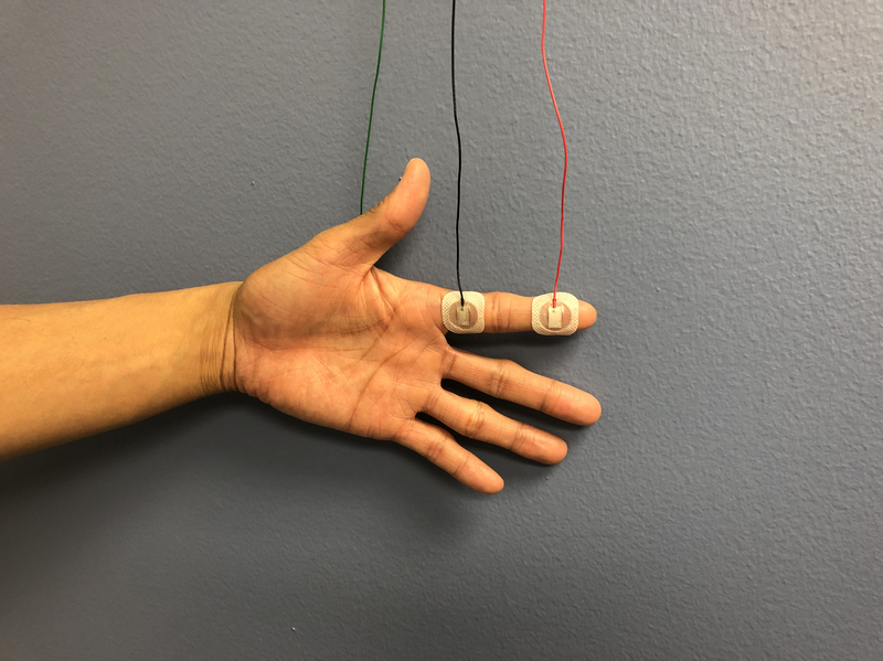
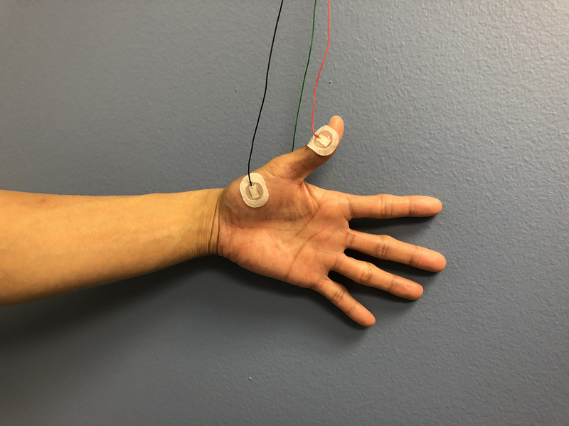
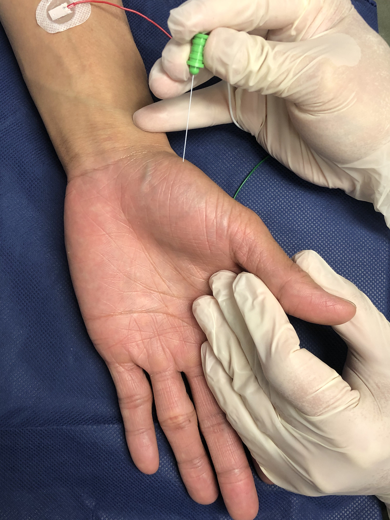
A. Median nerve sensory nerve conduction study setup. B. Median nerve motor nerve conduction study setup. C. Electromyography needle insertion into abductor pollicis brevis.
A. Median nerve sensory nerve conduction study setup. B. Median nerve motor nerve conduction study setup. C. Electromyography needle insertion into abductor pollicis brevis.
A. Median nerve sensory nerve conduction study setup. B. Median nerve motor nerve conduction study setup. C. Electromyography needle insertion into abductor pollicis brevis.
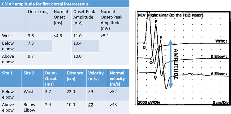
A. Motor nerve conduction study from the first dorsal interosseous muscle in a patient with mild cubital tunnel syndrome. Note the normal compound motor action potential (CMAP) amplitude levels are normal, but there is some slight slowing in the nerve conduction velocity across the elbow. B. Motor nerve conduction study from the abductor digiti minimi muscle in a patient with severe cubital tunnel syndrome. There is muscle wasting and loss of two-point discrimination on this patient’s clinical exam. Note the drastically decreased compound motor action potential (CMAP) amplitude levels in addition to marked slowing in the nerve conduction velocity across the elbow.
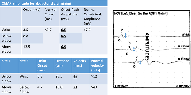
A. Motor nerve conduction study from the first dorsal interosseous muscle in a patient with mild cubital tunnel syndrome. Note the normal compound motor action potential (CMAP) amplitude levels are normal, but there is some slight slowing in the nerve conduction velocity across the elbow. B. Motor nerve conduction study from the abductor digiti minimi muscle in a patient with severe cubital tunnel syndrome. There is muscle wasting and loss of two-point discrimination on this patient’s clinical exam. Note the drastically decreased compound motor action potential (CMAP) amplitude levels in addition to marked slowing in the nerve conduction velocity across the elbow.
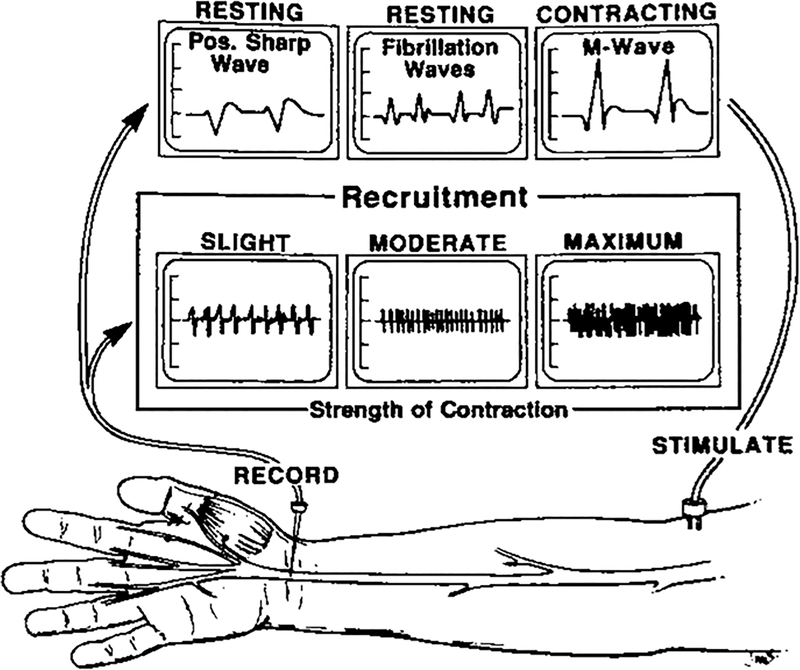
Waveforms seen during insertion, resting, and activation phases of EMG. (Reproduced with permission from Gelberman RH: Operative Nerve Repair and Reconstruction [Fig 10–2]; Ed: Gelberman RH, 1991. Lippincott)
References
-
- Nodera H, Herrmann DN, Holloway RG, Logigian EL. A Bayesian argument against rigid cut-offs in electrodiagnosis of median neuropathy at the wrist. Neurology. 2003. January;60(3):458–464. - PubMed
-
- Jablecki CK, Andary MT, Floeter MK, et al. Practice parameter: Electrodiagnostic studies in carpal tunnel syndrome. Report of the American Association of Electrodiagnostic Medicine, American Academy of Neurology, and the American Academy of Physical Medicine and Rehabilitation. Neurology. 2002. June;58(11):1589–92. - PubMed
-
- Pastare D, Therimadasamy AK, Lee E, Wilder-Smith EP. Sonography versus nerve conduction studies in patients referred with a clinical diagnosis of carpal tunnel syndrome. J Clin Ultrasound. 2009. September;37(7):389–93. - PubMed
-
- Robertson C, Saratsiotis J. A review of compressive ulnar neuropathy at the elbow. J Manipulative Physiol Ther. 2005. June;28(5):345. - PubMed
MeSH terms
Grants and funding
LinkOut - more resources
Full Text Sources
Other Literature Sources
