. 2021 Mar 19;17(3):e1009436.
doi: 10.1371/journal.ppat.1009436. eCollection 2021 Mar.

Resident bacteria contribute to opportunistic infections of the respiratory tract

Affiliations

Resident bacteria contribute to opportunistic infections of the respiratory tract

Yifan Wu et al. PLoS Pathog. .

Abstract

Opportunistic pathogens frequently cause volatile infections in hosts with compromised immune systems or a disrupted normal microbiota. The commensalism of diverse microorganisms contributes to colonization resistance, which prevents the expansion of opportunistic pathogens. Following microbiota disruption, pathogens promptly adapt to altered niches and obtain growth advantages. Nevertheless, whether and how resident bacteria modulate the growth dynamics of invasive pathogens and the eventual outcome of such infections are still unclear. Here, we utilized birds as a model animal and observed a resident bacterium exacerbating the invasion of Avibacterium paragallinarum (previously Haemophilus paragallinarum) in the respiratory tract. We first found that negligibly abundant Staphylococcus chromogenes, rather than Staphylococcus aureus, played a dominant role in Av. paragallinarum-associated infectious coryza in poultry based on epidemic investigations and in vitro analyses. Furthermore, we determined that S. chromogenes not only directly provides the necessary nutrition factor nicotinamide adenine dinucleotide (NAD+) but also accelerates its biosynthesis and release from host cells to promote the survival and growth of Av. paragallinarum. Last, we successfully intervened in Av. paragallinarum-associated infections in animal models using antibiotics that specifically target S. chromogenes. Our findings show that opportunistic pathogens can hijack commensal bacteria to initiate infection and expansion and suggest a new paradigm to ameliorate opportunistic infections by modulating the dynamics of resident bacteria.


Conflict of interest statement

The authors have declared that no competing interests exist.

Figures

Fig 1
Fig 1. S. chromogenes is closely related to Av. paragallinarum infection.
(A) Bacterial samples were collected from the nasal cavity and infraorbital sinuses of 20 birds with clinical symptoms. (B) Distribution of Staphylococcus spp. (C) Representative image of the satellitism of Av. paragallinarum on blood agar plates after cocultivation for 48 h. (D) Custom synergistic indexes, including the isolation rates (0–1), colocalization rates (0–1), and the mean radii of satellitism (cm).
Fig 2
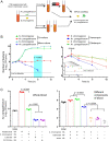
Fig 2. S. chromogenes supports the survival of Av. paragallinarum in vitro.
(A) Workflow of in vitro coculture experiments. S. chromogenes SC10 and Av. paragallinarum X1-1S-1 were cocultured or monocultured in homemade broth media with or without antimicrobial agents. The CFUs/mL bacteria of all groups were subsequently calculated. (B) The growth dynamic of S. chromogenes and Av. paragallinarum in the presence of 5% (v/v) whole blood with (picture on the right) or without antimicrobial agents (picture on the left). LOD, limit of detection. P values were determined by unpaired t-test. The values of three biological replicates are shown as individual points (n = 3). (C) Replication of Av. paragallinarum in the presence of whole blood or blood components. NA, not applicable. P values were determined by one-way ANOVA. The mean of three biological replicates is shown, and error bars represent the standard deviation (SD) (n  =  3).
Fig 3
Fig 3. S. chromogenes supports the growth of H. parasuis in vitro.
(A) Satellitism formed by S. chromogenes and H. parasuis after coculturing for 48 h. (B) H. parasuis displayed high density in the presence of S. chromogenes, together with either blood or serum. H. parasuis was monocultured or cocultured with S. chromogenes in TSB supplemented with different nutrients for 6 h. NA, not applicable. P values were determined by unpaired t-test. The mean of three biological replicates is shown, and error bars represent the standard deviation (SD) (n  =  3).
Fig 4
Fig 4. Accumulation of Staphylococcus-derived NAD+ promotes the survival of Av. paragallinarum.
(A) The NAD+ content in the medium. S. chromogenes SC10 was monocultured in TSB supplemented with 5% (v/v) serum at 37°C. (B) NAD+ content in cultures of five S. chromogenes isolates after 4 h. (C) NAD+ content in cultures of different Staphylococcus isolates after 4 h at 37°C. P values were determined by one-way ANOVA. The mean of three biological replicates is shown, and error bars represent the standard deviation (SD) (n  =  3).
Fig 5
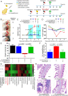
Fig 5. S. chromogenes aggravates the infection of Av. paragallinarum.
(A) Scheme of animal models. SPF white leghorn chickens were randomly divided into four groups with six chickens in each group. Bacterial infection (109 CFUs per bird), antimicrobial treatment, evaluation of food and water intake, and observation of clinical symptoms were all completed in the morning of each day. (B) Infection scores of symptoms. (C) S. chromogenes aggravated the clinical symptoms of birds infected with Av. paragallinarum. The difference was compared among the vancomycin treatment group and mono- or coinfection groups. (D) The food intake of each group was recorded every morning before renewal. (E) Body weight was recorded pre- and post-trial, and the weight obtained was calculated using subtraction. (F) Bacterial burden of the nasal cavity and infraorbital sinuses. (G) The bacterial community of the nasal cavity and infraorbital sinuses. (H) Serious tissue damage was observed in birds coinfected with S. chromogenes and Av. paragallinarum. A large number of inflammatory cells infiltrated the epithelial tissue or were contained in the respiratory secretions (a). Epithelial cells were damaged and shed (b). Scale bar = 100 μm. P values were determined by unpaired t-test. The mean of six biological replicates is shown, and error bars represent the standard deviation (SD) (n  =  6).
Fig 6
Fig 6. S. chromogenes hijacks host cells to promote the growth of Av. paragallinarum.
(A) S. chromogenes increased the number of extracellular Av. paragallinarum. MH-S cells were infected with S. chromogenes SC10 and Av. paragallinarum X1-1S-1 (MOI = 1) and treated with 10 μg/mL vancomycin for 6 h. (B) S. chromogenes promoted NAD+ release from the host cells. MH-S cells were treated the same as in (A). (C) S. chromogenes seriously damaged the membrane of mammalian cells. MH-S and A549 cells were infected with S. chromogenes SC10 (MOI = 1) for 6 h, and erythrocytes were coincubated with S. chromogenes (106 CFUs/mL) in PBS for 6 h. (D) The mRNA expression of NAD+ synthetases. MH-S cells were infected with S. chromogenes SC10 and/or Av. paragallinarum X1-1S-1 (MOI = 1) or incubated with 10 μg/mL vancomycin for 6 h. (E) S. chromogenes promoted the expression of NAMPT. A549 cells were infected with S. chromogenes SC10 and/or Av. paragallinarum X1-1S-1 (MOI = 1) for 6 h. (F) The NAMPT inhibitor (E)-daporinad reduced the number of Av. paragallinarum. MH-S cells were infected with S. chromogenes SC10 and Av. paragallinarum X1-1S-1 (MOI = 1) and treated with 1 μM (E)-daporinad and/or 10 μg/mL exogenous NAD+ for 6 h. (G, H) S. chromogenes increased the content of extracellular or intracellular NAD+. MH-S cells were infected with S. chromogenes (MOI = 1) and/or treated with 1 μM (E)-daporinad for 6 h. P values were determined by one-way ANOVA (A, B, D and E) and unpaired t-test (C, F, G and H). The mean of three biological replicates is shown, and error bars represent the standard deviation (SD) (n  =  3).
Fig 7
Fig 7. Resident bacteria promote the replication of Av. paragallinarum.
Resident bacteria such as S. chromogenes enhance NAD+ biosynthesis and release by host cells in the respiratory tract. Consequently, the increased NAD+ level in the extracellular environment accelerates the replication of Av. paragallinarum.

Similar articles

Cited by

References

    1. Kamada N, Nunez G. Regulation of the immune system by the resident intestinal bacteria. Gastroenterology. 2014;146(6):1477–88. 10.1053/j.gastro.2014.01.060 - DOI - PMC - PubMed
    1. Man WH, de Steenhuijsen Piters WA, Bogaert D. The microbiota of the respiratory tract: gatekeeper to respiratory health. Nat Rev Microbiol. 2017;15(5):259–70. 10.1038/nrmicro.2017.14 - DOI - PMC - PubMed
    1. Baumler AJ, Sperandio V. Interactions between the microbiota and pathogenic bacteria in the gut. Nature. 2016;535(7610):85–93. 10.1038/nature18849 - DOI - PMC - PubMed
    1. Perez-Lopez A, Behnsen J, Nuccio S-P, Raffatellu M. Mucosal immunity to pathogenic intestinal bacteria. Nat Rev Immunol. 2016;16(3):135–48. 10.1038/nri.2015.17 - DOI - PubMed
    1. Cohen TS, Hilliard JJ, Jones-Nelson O, Keller AE, O’Day T, Tkaczyk C, et al.. Staphylococcus aureus alpha toxin potentiates opportunistic bacterial lung infections. Sci Transl Med. 2016;8(329):329ra31. 10.1126/scitranslmed.aad9922 - DOI - PubMed

Publication types

MeSH terms

Substances

Supplementary concepts

LinkOut - more resources