. 2021 Mar 19;16(3):e0249020.
doi: 10.1371/journal.pone.0249020. eCollection 2021.

Tracking down the White Plague. Chapter three: Revision of endocranial abnormally pronounced digital impressions as paleopathological diagnostic criteria for tuberculous meningitis

Affiliations

Tracking down the White Plague. Chapter three: Revision of endocranial abnormally pronounced digital impressions as paleopathological diagnostic criteria for tuberculous meningitis

Olga Spekker et al. PLoS One. .

Abstract

Abnormally pronounced digital impressions (APDIs) on the endocranial surface develop secondary to a prolonged rise in the intracranial pressure. This can result from a number of pathological conditions, including hydrocephalus due to tuberculous meningitis (TBM). APDIs have been described with relation to TBM not only in the modern medical literature but also in several paleopathological studies. However, APDIs are not pathognomonic for TBM and their diagnostic value for identifying TBM in past human populations has not been evaluated in identified pre-antibiotic era skeletons. To assess the diagnostic value of APDIs for the first time, a macroscopic investigation was performed on skeletons from the Terry Collection (Smithsonian Institution, Washington, DC, USA). Our material consisted of 234 skeletons with tuberculosis (TB) as the cause of death (TB group) and 193 skeletons with non-tuberculous (NTB) causes of death (NTB group). The macroscopic examination focused on the stage of the prominence and frequency of APDIs in the TB group and NTB group. To determine the significance of difference (if any) in the frequency of APDIs between the two groups, χ2 testing of our data was conducted. We found that APDIs were twice as common in the TB group than in the NTB group. The χ2 comparison of the frequencies of APDIs revealed a statistically significant difference between the two groups. In addition, APDIs with more pronounced stages were recorded more frequently in the TB group. Our results indicate that APDIs can be considered as diagnostic criteria for TBM in the paleopathological practice. With suitable circumspection, their utilization provides paleopathologists with a stronger basis for identifying TB and consequently, with a more sensitive means of assessing TB frequency in past human populations.


Conflict of interest statement

The authors have declared that no competing interests exist.

Figures
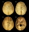
Fig 1
Fig 1. Reference cases selected for the classification of individuals exhibiting APDIs on the endocranial surface of the skull.
A) Very slight stage represented by shallow DIs predominantly localized over the anterior portion of the inner skull surface (Terry No. 30R: 26-year-old, male, died of TBM–skullcap); B) Slight stage represented by deeper DIs particularly situated over the anterior and middle portions of the endocranial surface (Terry No. 382R: 26-year-old, male, died of pulmonary TB–skullcap); C) Pronounced stage represented by deep DIs localized all over the inner skull surface (Terry No. 1033: 26-year-old, male, died of pulmonary TB–skullcap); and D) Pronounced stage represented by deep DIs situated all over the endocranial surface (Terry No. 1033: 26-year-old, male, died of pulmonary TB–skull base).
Fig 2
Fig 2. Different stages of the prominence of APDIs (white arrows) on the inner surface of the squamous part of the frontal bone.
A) Very slight stage represented by shallow DIs on the endocranial surface of the frontal bone of Terry No. 1036 (38-year-old, male, died of pulmonary TB); B) Slight stage represented by deeper DIs on the inner skull surface of the frontal bone of Terry No. 265 (32-year-old, male, died of TB); and C) Pronounced stage represented by deep DIs on the endocranial surface of the frontal bone of Terry No. 251 (34-year-old, male, died of pulmonary TB).
Fig 3
Fig 3. Distribution of the selected individuals from the Terry Collection by the presence of APDIs (considering the different stages of the prominence of lesions).
A) Distribution of individuals who died of TB; and B) Distribution of individuals who died of NTB causes. (In both the TB group (A) and NTB group (B), the larger pie chart shows the distribution of all individuals by the presence of APDIs (i.e., not present (white) or present (dark blue)). Whereas, the smaller pie chart shows the distribution of individuals displaying APDIs by the prominence stage of the presented APDIs (i.e., very slight (light blue), slight (orange), and pronounced (yellow)). The percentage values were calculated with respect to the total number of individuals in both the TB group (NTB = 234) and NTB group (NNTB = 193)).
Fig 4
Fig 4. Distribution of the selected individuals from the Terry Collection by the presence of APDIs (considering the co-occurrence of APDIs with other probable TBM-related endocranial lesion types and/or with possible TB-associated non-endocranial lesion types).
A) Distribution of individuals who died of TB; and B) Distribution of individuals who died of NTB causes. (In both the TB group (A) and NTB group (B), the larger pie chart shows the distribution of all individuals by the presence of APDIs (i.e., not present (white), present but alone (black) or present in association with other likely TB-related lesion(s) (dark blue)). The smaller pie chart shows the distribution of individuals displaying APDIs in association with other probable TB-related lesion(s) by the type of the lesion(s) simultaneously occurring with APDIs (i.e., besides APDIs, only other probable TBM-related endocranial lesion(s) are present (yellow), besides APDIs, only possible TB-associated non-endocranial lesion(s) are present (orange) or besides APDIs, both other probable TBM-related endocranial lesion(s) and possible TB-associated non-endocranial lesion(s) are present (light blue)). The percentage values were calculated with respect to the total number of individuals in both the TB group (NTB = 234) and NTB group (NNTB = 193)).

Similar articles

Cited by

References

    1. Connolly CA, Gibson ME. The “White Plague” and color: Children, race, and tuberculosis in Virginia 1900–1935. J Pediatr Nurs. 2011;26(3): 230–238. 10.1016/j.pedn.2010.01.011 - DOI - PMC - PubMed
    1. Gagneux S. Host-pathogen co-evolution in human tuberculosis. Phil Trans R Soc B 2012;367(1590): 850–859. 10.1098/rstb.2011.0316 - DOI - PMC - PubMed
    1. Galagan JE. Genomic insights into tuberculosis. Nat Rev Genet. 2014;15(5): 307–320. 10.1038/nrg3664 - DOI - PubMed
    1. Kelley MA, Micozzi MS. Rib lesions in chronic pulmonary tuberculosis. Am J Phys Anthropol. 1984;65(4): 381–386. 10.1002/ajpa.1330650407 - DOI - PubMed
    1. Roberts CA, Lucy D, Manchester K. Inflammatory lesions of ribs: An analysis of the Terry Collection. Am J Phys Anthropol. 1994;95(2): 169–182. 10.1002/ajpa.1330950205 - DOI - PubMed

Publication types

LinkOut - more resources