

Tubulin acetylation increases cytoskeletal stiffness to regulate mechanotransduction in striated muscle
- PMID: 33740038
- PMCID: PMC7988512
- DOI: 10.1085/jgp.202012743
Tubulin acetylation increases cytoskeletal stiffness to regulate mechanotransduction in striated muscle
Abstract
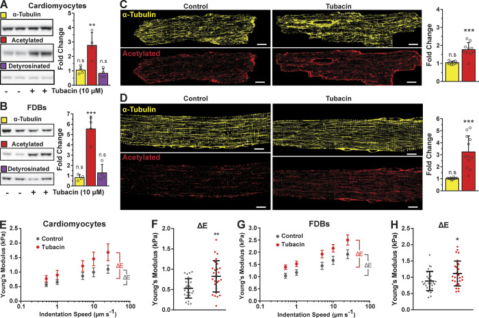
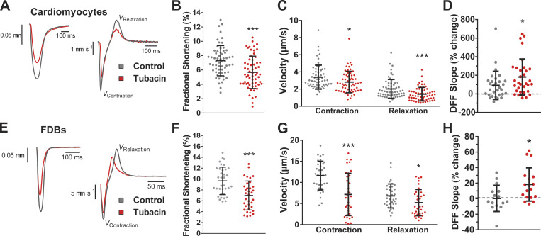
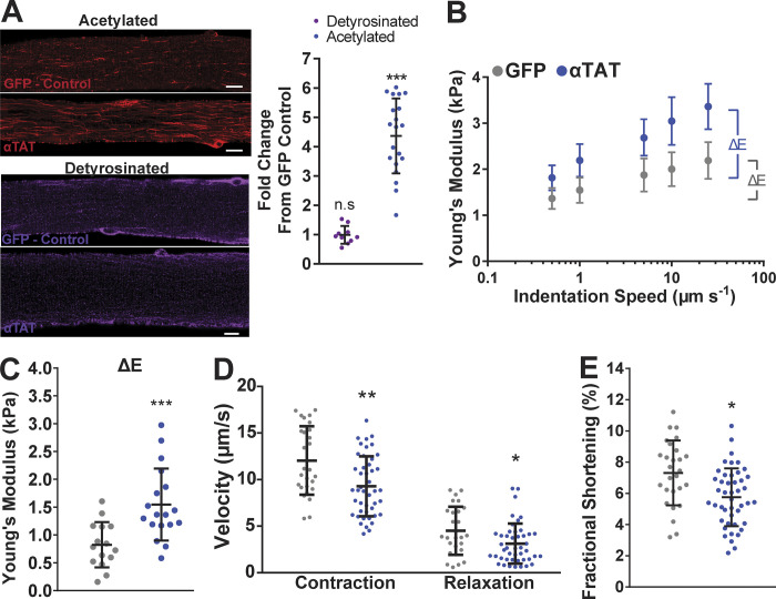
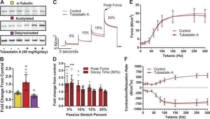
Microtubules tune cytoskeletal stiffness, which affects cytoskeletal mechanics and mechanotransduction of striated muscle. While recent evidence suggests that microtubules enriched in detyrosinated α-tubulin regulate these processes in healthy muscle and increase them in disease, the possible contribution from several other α-tubulin modifications has not been investigated. Here, we used genetic and pharmacologic strategies in isolated cardiomyocytes and skeletal myofibers to increase the level of acetylated α-tubulin without altering the level of detyrosinated α-tubulin. We show that microtubules enriched in acetylated α-tubulin increase cytoskeletal stiffness and viscoelastic resistance. These changes slow rates of contraction and relaxation during unloaded contraction and increased activation of NADPH oxidase 2 (Nox2) by mechanotransduction. Together, these findings add to growing evidence that microtubules contribute to the mechanobiology of striated muscle in health and disease.
© 2021 Coleman et al.
Figures
Similar articles
-
Relationship between the Golgi complex and microtubules enriched in detyrosinated or acetylated alpha-tubulin: studies on cells recovering from nocodazole and cells in the terminal phase of cytokinesis.Cell Tissue Res. 1993 Sep;273(3):457-66. doi: 10.1007/BF00333700. Cell Tissue Res. 1993. PMID: 8402828
-
Posttranslational modifications of alpha tubulin: detyrosination and acetylation differentiate populations of interphase microtubules in cultured cells.J Cell Biol. 1988 Apr;106(4):1213-20. doi: 10.1083/jcb.106.4.1213. J Cell Biol. 1988. PMID: 3283150 Free PMC article.
-
Posttranslational modifications of tubulin in teleost photoreceptor cytoskeletons.Cell Mol Neurobiol. 1991 Dec;11(6):593-610. doi: 10.1007/BF00741448. Cell Mol Neurobiol. 1991. PMID: 1782652 Free PMC article.
-
The tubulin code: molecular components, readout mechanisms, and functions.J Cell Biol. 2014 Aug 18;206(4):461-72. doi: 10.1083/jcb.201406055. J Cell Biol. 2014. PMID: 25135932 Free PMC article. Review.
-
[On the road to deciphering the tubulin code: focus on acetylation and detyrosination].Med Sci (Paris). 2018 Dec;34(12):1047-1055. doi: 10.1051/medsci/2018295. Epub 2019 Jan 9. Med Sci (Paris). 2018. PMID: 30623774 Review. French.
Cited by
-
Non-catalytic allostery in α-TAT1 by a phospho-switch drives dynamic microtubule acetylation.J Cell Biol. 2022 Nov 7;221(11):e202202100. doi: 10.1083/jcb.202202100. Epub 2022 Oct 12. J Cell Biol. 2022. PMID: 36222836 Free PMC article.
-
Histone deacetylase 6 inhibition promotes microtubule acetylation and facilitates autophagosome-lysosome fusion in dystrophin-deficient mdx mice.Acta Physiol (Oxf). 2025 Jan;241(1):e14243. doi: 10.1111/apha.14243. Epub 2024 Oct 18. Acta Physiol (Oxf). 2025. PMID: 39422111
-
TRPV4 modulates substrate stiffness mechanosensing and transcellular pore formation in human Schlemm's canal cells.bioRxiv [Preprint]. 2025 May 26:2025.05.25.656000. doi: 10.1101/2025.05.25.656000. bioRxiv. 2025. PMID: 40501826 Free PMC article. Preprint.
-
Ryanodine receptor stabilization therapy suppresses Ca2+- based arrhythmias in a novel model of metabolic HFpEF.J Mol Cell Cardiol. 2024 Oct;195:68-72. doi: 10.1016/j.yjmcc.2024.07.006. Epub 2024 Jul 23. J Mol Cell Cardiol. 2024. PMID: 39053573
-
Aging, Osteo-Sarcopenia, and Musculoskeletal Mechano-Transduction.Front Rehabil Sci. 2021;2:782848. doi: 10.3389/fresc.2021.782848. Epub 2021 Dec 6. Front Rehabil Sci. 2021. PMID: 36004321 Free PMC article.
References
-
- Belanto, J.J., Olthoff J.T., Mader T.L., Chamberlain C.M., Nelson D.M., McCourt P.M., Talsness D.M., Gundersen G.G., Lowe D.A., and Ervasti J.M.. 2016. Independent variability of microtubule perturbations associated with dystrophinopathy. Hum. Mol. Genet. 25:4951–4961. 10.1093/hmg/ddw318 - DOI - PMC - PubMed
-
- Brynnel, A., Hernandez Y., Kiss B., Lindqvist J., Adler M., Kolb J., van der Pijl R., Gohlke J., Strom J., Smith J., et al. . 2018. Downsizing the molecular spring of the giant protein titin reveals that skeletal muscle titin determines passive stiffness and drives longitudinal hypertrophy. eLife. 7:e40532. 10.7554/eLife.40532 - DOI - PMC - PubMed
-
- Burkholder, T.J., and Lieber R.L.. 2001. Sarcomere length operating range of vertebrate muscles during movement. J. Exp. Biol. 204:1529–1536. - PubMed
-
- Chen, C.Y., Caporizzo M.A., Bedi K., Vite A., Bogush A.I., Robison P., Heffler J.G., Salomon A.K., Kelly N.A., Babu A., et al. . 2018. Suppression of detyrosinated microtubules improves cardiomyocyte function in human heart failure. Nat. Med. 24:1225–1233. 10.1038/s41591-018-0046-2 - DOI - PMC - PubMed
Publication types
MeSH terms
Substances
Grants and funding
LinkOut - more resources
Full Text Sources
Other Literature Sources
Miscellaneous
