. 2021 Sep;31(9):7132-7142.
doi: 10.1007/s00330-021-07809-w. Epub 2021 Mar 19.

Reduced-iodine-dose dual-energy coronary CT angiography: qualitative and quantitative comparison between virtual monochromatic and polychromatic CT images

Affiliations

Reduced-iodine-dose dual-energy coronary CT angiography: qualitative and quantitative comparison between virtual monochromatic and polychromatic CT images

David C Rotzinger et al. Eur Radiol. 2021 Sep.

Abstract

Objectives: To quantitatively evaluate the impact of virtual monochromatic images (VMI) on reduced-iodine-dose dual-energy coronary computed tomography angiography (CCTA) in terms of coronary lumen segmentation in vitro, and secondly to assess the image quality in vivo, compared with conventional CT obtained with regular iodine dose.

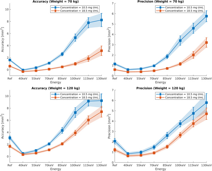
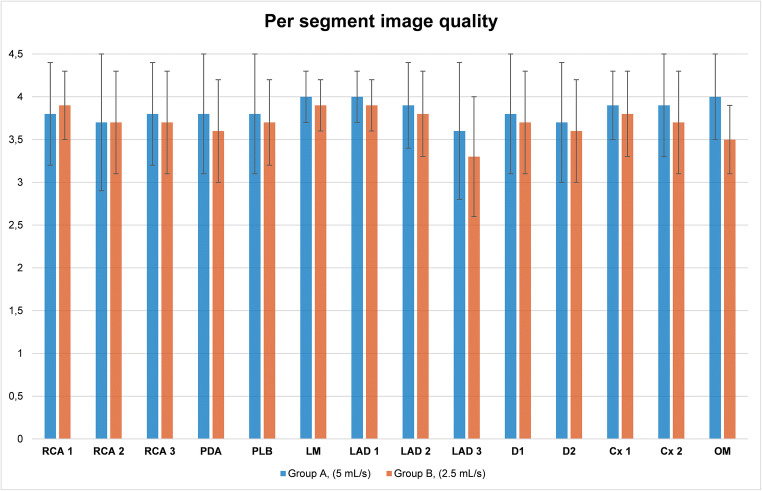
Materials and methods: A phantom simulating regular and reduced iodine injection was used to determine the accuracy and precision of lumen area segmentation for various VMI energy levels. We retrospectively included 203 patients from December 2017 to August 2018 (mean age, 51.7 ± 16.8 years) who underwent CCTA using either standard (group A, n = 103) or reduced (group B, n = 100) iodine doses. Conventional images (group A) were qualitatively and quantitatively compared with 55-keV VMI (group B). We recorded the location of venous catheters.

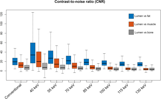
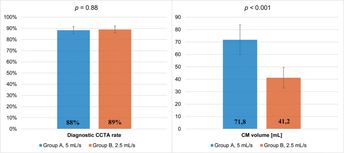
Results: In vitro, VMI outperformed conventional CT, with a segmentation accuracy of 0.998 vs. 1.684 mm2, respectively (p < 0.001), and a precision of 0.982 vs. 1.229 mm2, respectively (p < 0.001), in simulated overweight adult subjects. In vivo, the rate of diagnostic CCTA in groups A and B was 88.4% (n = 91/103) vs. 89% (n = 89/100), respectively, and noninferiority of protocol B was inferred. Contrast-to-noise ratios (CNR) of lumen versus fat and muscle were higher in group B (p < 0.001) and comparable for lumen versus calcium (p = 0.423). Venous catheters were more often placed on the forearm or hand in group B (p < 0.001).

Conclusion: In vitro, low-keV VMI improve vessel area segmentation. In vivo, low-keV VMI allows for a 40% iodine dose and injection rate reduction while maintaining diagnostic image quality and improves the CNR between lumen versus fat and muscle.

Key points: • Dual-energy coronary CT angiography is becoming increasingly available and might help improve patient management. • Compared with regular-iodine-dose coronary CT angiography, reduced-iodine-dose dual-energy CT with low-keV monochromatic image reconstructions performed better in phantom-based vessel cross-sectional segmentation and proved to be noninferior in vivo. • Patients receiving reduced-iodine-dose dual-energy coronary CT angiography often had the venous catheter placed on the forearm or wrist without compromising image quality.

Keywords: Computed tomography angiography; Coronary vessels; Dimensional measurement accuracy; Iodine; Phantoms, imaging.


Conflict of interest statement

The authors of this manuscript declare no relationships with any companies whose products or services may be related to the subject matter of the article.

Figures

Fig. 1
Fig. 1
In vitro accuracy and precision of vessel lumen segmentation as a function of iodine dose (10.5 or 18.5 mg/mL) and patient size (70 or 120 kg). The conventional reconstruction “Ref” at 18.5 mL approximates protocol A in the clinical study, whereas the virtual monochromatic images (VMI) at 10.5 mg/mL approximate protocol B. Whatever the scanning condition, low VMI energies, up to a maximum of 55 keV, yielded peak segmentation accuracy and precision
Fig. 2
Fig. 2
Boxplots show contrast-to-noise ratios (CNR) for different scenarios: lumen vs. fat to approximate an epicardial vessel surrounded by fat, lumen vs. muscle to approximate a non-calcified plaque, and lumen vs. trabecular bone to approximate a calcified plaque. When comparing group A (5 mL/s) with group B (2.5 mL/s reconstructed at 55 keV), no significant difference in lumen to bone CNR was found, but group B had significantly better lumen to fat and lumen to muscle CNR
Fig. 3
Fig. 3
Coronary CT angiography obtained with 40 mL contrast medium injected at 2.5 mL/s and reconstructed as virtual monoenergetic images at 55 keV, depicting the LAD, LCx, and RCA (ac), in a 68-year-old male. Coronary CT angiography obtained with 70 mL contrast agent injected at 5 mL/s and reconstructed as conventional polychromatic images, depicting the LAD, LCx, and RCA (df), in a 48-year-old female. LAD, left anterior descending; Cx, circumflex artery; RCA, right coronary artery; keV, kilo-electronvolt
Fig. 4
Fig. 4
Proportion of interpretable CCTA (± standard error) and contrast agent volume (± standard deviation) in groups A and B. CM, contrast medium; CCTA, coronary computed tomography angiography. Conventional polychromatic images in group A and virtual monoenergetic images at 55 keV in group B
Fig. 5
Fig. 5
Per segment qualitative image analysis in group A and B and univariate comparison. Error bars represent standard deviation. Proximal right coronary artery (RCA 1), p = 0.96; mid right coronary artery (RCA 2), p = 0.552; distal right coronary artery (RCA 3), p = 0.216; posterior descending artery (PDA), p = 0.001; posterolateral branch (PLB), p = 0.010; left main (LM), p = 0.05; proximal left anterior descending (LAD 1), p = 0.004; mid left anterior descending (LAD 2), p = 0.023; distal left anterior descending (LAD 3), p = 0.001; first diagonal (D1), p = 0.031; second diagonal (D2), p = 0.059, proximal circumflex (Cx 1), p = 0.009; distal circumflex (Cx 2), p < 0.001; obtuse marginal (OM), p = 0.003. LAD, left anterior descending; LCx, left cirumflex; LM, left main; OM, obtuse marginal; PDA, posterior descending artery; PLB, posterolateral branch; RCA, right coronary artery

References

    1. Schoepf UJ, Zwerner PL, Savino G, Herzog C, Kerl JM, Costello P. Coronary CT angiography. Radiology. 2007;244:48–63. doi: 10.1148/radiol.2441052145. - DOI - PubMed
    1. Brown JR, Robb JF, Block CA, et al. Does safe dosing of iodinated contrast prevent contrast-induced acute kidney injury? Circ Cardiovasc Interv. 2010;3:346–350. doi: 10.1161/CIRCINTERVENTIONS.109.910638. - DOI - PMC - PubMed
    1. Van Cauteren T, Honoria Da Silva E, Van Gompel G, et al. Iodine dose of administered contrast media affects the level of radiation-induced DNA damage during cardiac CT scans. AJR Am J Roentgenol. 2019;213:404–409. doi: 10.2214/AJR.18.20950. - DOI - PubMed
    1. American College of Radiology (2020) ACR manual on contrast media v10.3. Available via https://www.acr.org/-/media/ACR/Files/Clinical-Resources/Contrast_Media.pdf. Accessed Apr 18 2020
    1. van Hamersvelt RW, Eijsvoogel NG, Mihl C, et al. Contrast agent concentration optimization in CTA using low tube voltage and dual-energy CT in multiple vendors: a phantom study. Int J Cardiovasc Imaging. 2018;34:1265–1275. doi: 10.1007/s10554-018-1329-x. - DOI - PubMed

LinkOut - more resources