. 2021 Jul;479(1):79-94.
doi: 10.1007/s00428-021-03038-0. Epub 2021 Mar 19.

Cardiac hypertrophy at autopsy

Affiliations

Cardiac hypertrophy at autopsy

Cristina Basso et al. Virchows Arch. 2021 Jul.

Erratum in

  • Correction to: Cardiac hypertrophy at autopsy.
    Basso C, Michaud K, d'Amati G, Banner J, Lucena J, Cunningham K, Leone O, Vink A, van der Wal AC, Sheppard MN; Association for European Cardiovascular Pathology. Basso C, et al. Virchows Arch. 2021 Jul;479(1):95. doi: 10.1007/s00428-021-03118-1. Virchows Arch. 2021. PMID: 33987695 Free PMC article. No abstract available.

Abstract

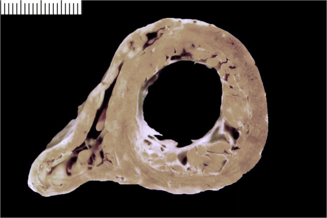
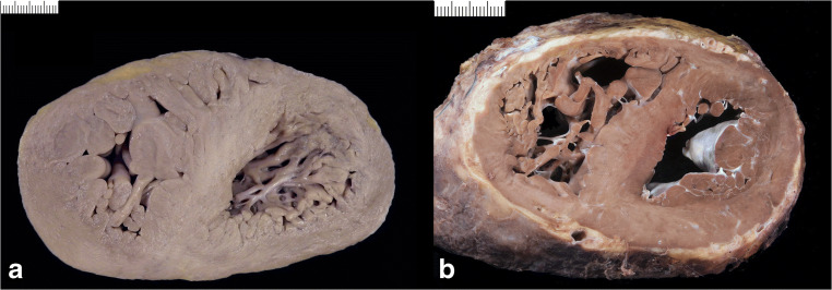
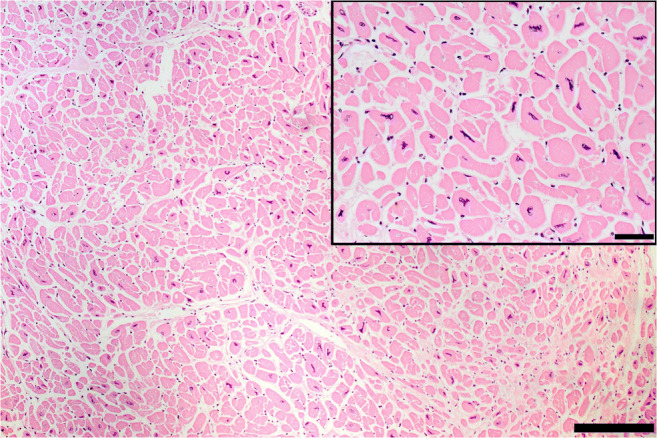
Since cardiac hypertrophy may be considered a cause of death at autopsy, its assessment requires a uniform approach. Common terminology and methodology to measure the heart weight, size, and thickness as well as a systematic use of cut off values for normality by age, gender, and body weight and height are needed. For these reasons, recommendations have been written on behalf of the Association for European Cardiovascular Pathology. The diagnostic work up implies the search for pressure and volume overload conditions, compensatory hypertrophy, storage and infiltrative disorders, and cardiomyopathies. Although some gross morphologic features can point to a specific diagnosis, systematic histologic analysis, followed by possible immunostaining and transmission electron microscopy, is essential for a final diagnosis. If the autopsy is carried out in a general or forensic pathology service without expertise in cardiovascular pathology, the entire heart (or pictures) together with mapped histologic slides should be sent for a second opinion to a pathologist with such an expertise. Indication for postmortem genetic testing should be integrated into the multidisciplinary management of sudden cardiac death.

Keywords: Autopsy; Cardiovascular diseases; Diagnostic criteria; Hypertrophy; Quality in pathology.


Conflict of interest statement

The authors declare no conflicts of interest.

Figures

Fig. 1
Fig. 1
a Diagrams illustrating the three cross sections of the hearts: the first mid-ventricular one and at least two additional parallel sections towards the apex. b On the left: left ventricular (posterior), septal (middle), and right ventricular (posterior) thickness measurements, by excluding trabeculae and papillary muscles. On the right: transverse size and longitudinal size measurements on the posterior aspect of the heart. ALMV, anterior leaflet mitral valve; ALPM, antero-lateral papillary muscle; IVC, inferior vena cava; LV, left ventricle; LVFW, left ventricular free wall; LSPV, left superior pulmonary vein; LIPV, left inferior pulmonary vein; PLMV, posterior leaflet mitral valve; PSPM, postero-septal papillary muscle; RIPV, right inferior pulmonary vein; RSPV, right superior pulmonary vein; RV, right ventricle; RVFW, right ventricular free wall; SVC, superior vena cava; VS, ventricular septum
Fig. 2
Fig. 2
Normal heart vs. cardiac hypertrophy. a Cross section of a normal heart in a perinatal death: the right ventricular free wall thickness is 3.5 mm, the left ventricular is 5 mm, and the septum 5.5 mm. b Cross section of a normal adult heart: the right ventricular free wall thickness is 2 mm, the left ventricular is 12 mm, and the septum 13 mm. c Cross section of hypertrophic heart in an adult: the right ventricular free wall thickness is 7 mm, the left ventricular is 21 mm, and the septum 22 mm. d Histology of a showing hypercellularity which is normal for a perinatal myocardium (high number of cardiac myocyte/myocardial area) (bar = 100 micron). e Histology of b with diameter of cardiac myocytes within normal values (mean diameter 12 micron) (bar = 100 micron). f Histology of c with cardiac myocyte hypertrophy (mean diameter 20 micron) (bar = 100 micron)
Fig. 3
Fig. 3
Diagrams illustrating various patterns of cardiac hypertrophy. a Symmetric, concentric. b Symmetric, eccentric. c Asymmetric septal. d Asymmetric antero-lateral. e Biventricular. f Right ventricular
Fig. 4
Fig. 4
Cardiac hypertrophy. a Concentric type (with increased LV wall thickness). b Eccentric type (with increased LV cavity)
Fig. 5
Fig. 5
“Primary” cardiomyopathies. a Asymmetric septal hypertrophic cardiomyopathy with septal/LV free wall thickness ratio >1.3. b Asymmetric antero-lateral hypertrophic cardiomyopathy. c Dilated cardiomyopathy with biventricular dilatation
Fig. 6
Fig. 6
“Secondary” cardiomyopathies. a Concentric biventricular hypertrophy in storage disease/glycogenosis. d Histology of a showing diffuse cardiac myocytes vacuolization (hematoxylin eosin stain bar = 100 micron; insert TEM with intracellular glycogen deposits). b Concentric biventricular hypertrophy in storage disease/Fabry disease. e Histology of b showing extensive vacuolization of the cardiac myocytes (hematoxylin eosin stain bar = 100 micron; insert TEM with concentric lamellar bodies). e Concentric biventricular hypertrophy in infiltrative disease/amyloidosis. f Histology of c with amorphous material deposition in the interstitial space (hematoxylin eosin stain bar = 100 micron; insert shows the amorphous Congo red–positive substance at polarized light)
Fig. 7
Fig. 7
Cardiac hypertrophy due to pressure overload. a Hypertensive heart disease. b Aortic valve stenosis due to dystrophic calcification
Fig. 8
Fig. 8
Compensatory hypertrophy. a Chronic ischemic heart disease with previous lateral myocardial infarction. b Subacute-chronic myocarditis with mild left ventricular hypertrophy
Fig. 9
Fig. 9
Left ventricular noncompaction cardiomyopathy/dilated. Note the left ventricular cavity enlargement, thinning of the compact layer with prominent trabeculae of the left ventricular free wall. Note the endocardial fibroelastosis following the outline of the trabeculae
Fig. 10
Fig. 10
Right ventricular hypertrophy. a Primary pulmonary hypertension. b Congenital heart disease (surgically repaired atrio-ventricular septal defect)
Fig. 11
Fig. 11
Histology of cardiac myocyte hypertrophy. Note the increased cardiac myocyte diameter and the bizarre, irregular, and hyperchromatic nuclei (hematoxylin eosin stain bar = 50 micron). A close-up (bar = 100 micron) is seen in the insert
Fig. 12
Fig. 12
Histology of hypertrophic cardiomyopathy. a Myocyte bundle disarray (hematoxylin eosin stain). b Myocyte bundle disarray (trichrome stain). c Interstitial fibrosis (trichrome stain). d Replacement-type fibrosis with small vessel disease (trichrome stain). All bar = 100 micron

Similar articles

Cited by

References

    1. Cunningham KS, Spears DA, Care M. Evaluation of cardiac hypertrophy in the setting of sudden cardiac death. Forensic Sci Res. 2019;4(3):223–240. doi: 10.1080/20961790.2019.1633761. - DOI - PMC - PubMed
    1. Basso C, Aguilera B, Banner J, Cohle S, d'Amati G, de Gouveia RH, di Gioia C, Fabre A, Gallagher PJ, Leone O, Lucena J, Mitrofanova L, Molina P, Parsons S, Rizzo S, Sheppard MN, Mier MPS, Suvarna SK, Thiene G, van der Wal A, Vink A, Michaud K, Association for European Cardiovascular Pathology Guidelines for autopsy investigation of sudden cardiac death: 2017 update from the Association for European Cardiovascular Pathology. Virchows Arch. 2017;471(6):691–705. doi: 10.1007/s00428-017-2221-0. - DOI - PMC - PubMed
    1. Elliott PM, Anastasakis A, Borger MA, Borggrefe M, Cecchi F, Charron P, Hagege AA, Lafont A, Limongelli G, Mahrholdt H, McKenna WJ, Mogensen J, Nihoyannopoulos P, Nistri S, Pieper PG, Pieske B, Rapezzi C, Rutten FH, Tillmanns C, Watkins H. ESC Guidelines on diagnosis and management of hypertrophic cardiomyopathy: the Task Force for the Diagnosis and Management of Hypertrophic Cardiomyopathy of the European Society of Cardiology (ESC) Eur Heart J. 2014;35(39):2733–2779. doi: 10.1093/eurheartj/ehu284. - DOI - PubMed
    1. Maron BJ, Towbin JA, Thiene G, Antzelevitch C, Corrado D, Arnett D, Moss AJ, Seidman CE, Young JB, American Heart Association. Council on Clinical Cardiology, Heart Failure and Transplantation Committee. Quality of Care and Outcomes Research and Functional Genomics and Translational Biology Interdisciplinary Working Groups. Council on Epidemiology and Prevention Contemporary definitions and classification of the cardiomyopathies: an American Heart Association Scientific Statement from the Council on Clinical Cardiology, Heart Failure and Transplantation Committee; Quality of Care and Outcomes Research and Functional Genomics and Translational Biology Interdisciplinary Working Groups; and Council on Epidemiology and Prevention. Circulation. 2006;113(14):1807–1816. doi: 10.1161/CIRCULATIONAHA.106.174287. - DOI - PubMed
    1. Elliott P, Andersson B, Arbustini E, Bilinska Z, Cecchi F, Charron P, Dubourg O, Kühl U, Maisch B, McKenna WJ, Monserrat L, Pankuweit S, Rapezzi C, Seferovic P, Tavazzi L, Keren A. Classification of the cardiomyopathies: a position statement from the European Society Of Cardiology Working Group on Myocardial and Pericardial Diseases. Eur Heart J. 2008;29(2):270–276. doi: 10.1093/eurheartj/ehm342. - DOI - PubMed

LinkOut - more resources