. 2021 Mar;1(3):e35.
doi: 10.1002/cpz1.35.

Isolating and Analyzing Protein Containing Granules from Cells

Affiliations

Isolating and Analyzing Protein Containing Granules from Cells

Rachel A Victor et al. Curr Protoc. 2021 Mar.

Erratum in

Abstract

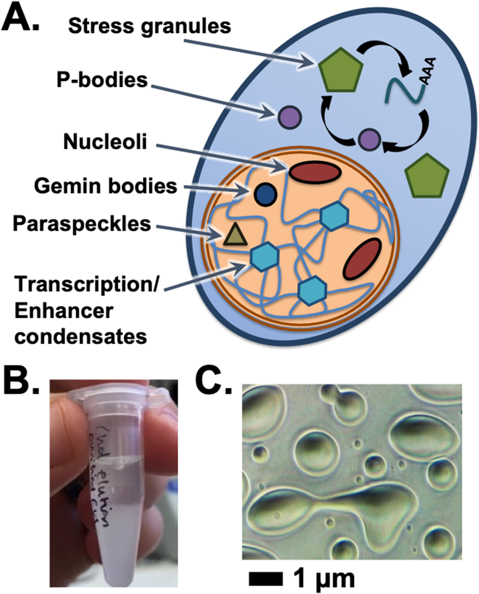
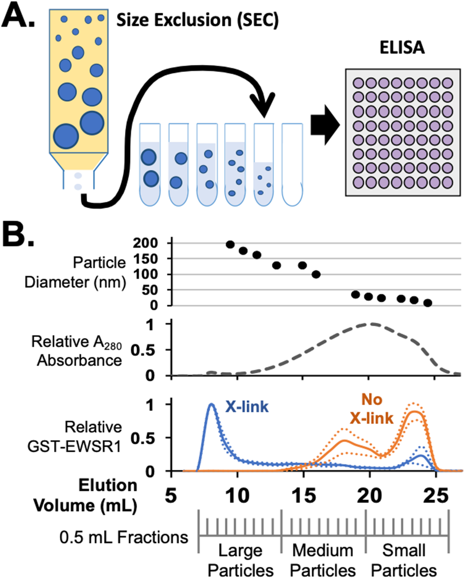
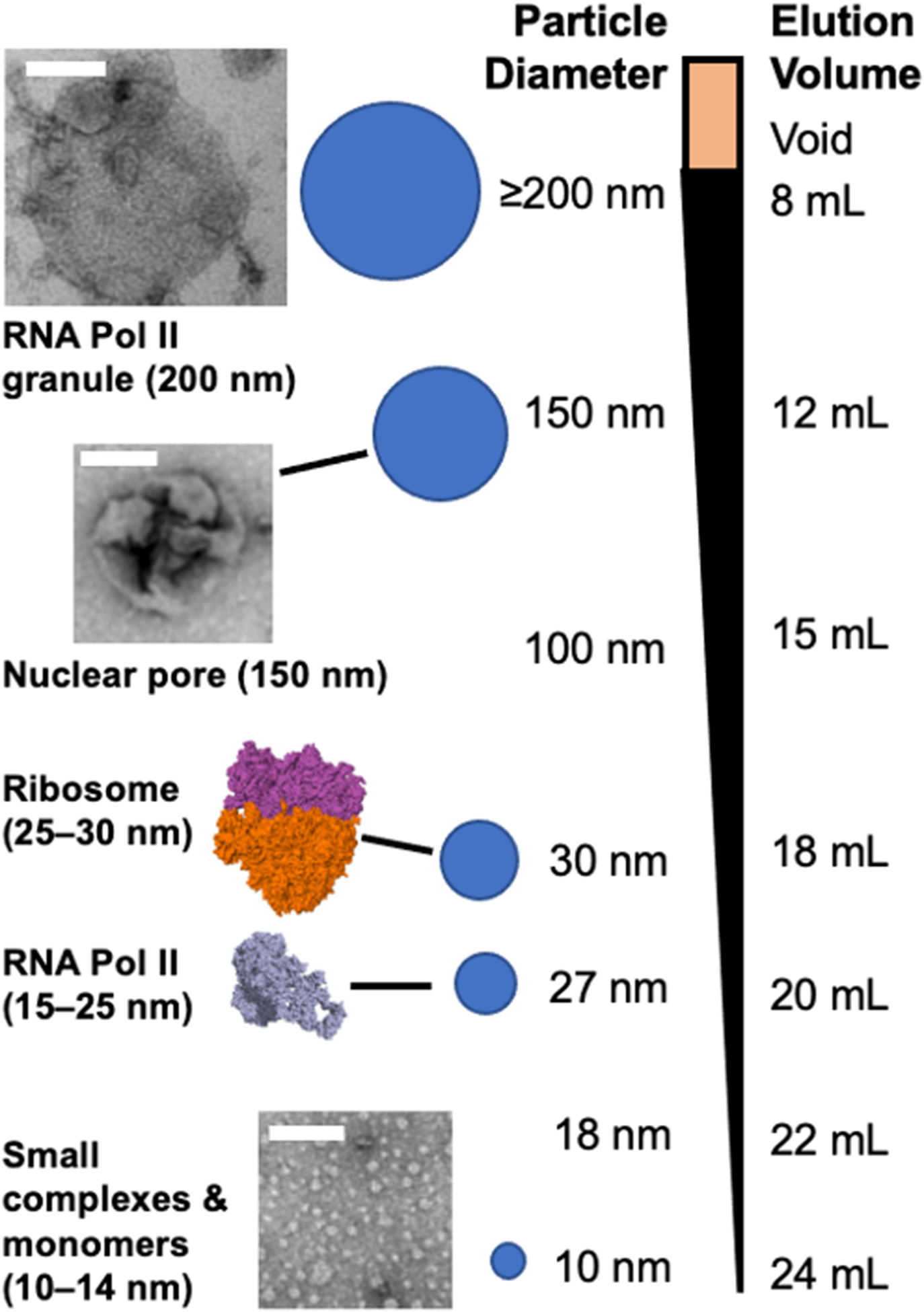
Recent advancements in detection methods have made protein condensates, also called granules, a major area of study, but tools to characterize these assemblies need continued development to keep up with evolving paradigms. We have optimized a protocol to separate condensates from cells using chemical cross-linking followed by size-exclusion chromatography (SEC). After SEC fractionation, the samples can be characterized by a variety of approaches including enzyme-linked immunosorbent assay, dynamic light scattering, electron microscopy, and mass spectrometry. The protocol described here has been optimized for cultured mammalian cells and E. coli expressing recombinant proteins. Since the lysates are fractionated by size, this protocol can be modified to study other large protein assemblies, including the nuclear pore complex, and for other tissues or organisms. © 2021 Wiley Periodicals LLC. Basic Protocol 1: SEC separation of cross-linked mammalian cell lysates Alternate Protocol: Preparation of non-crosslinked mammalian cells Basic Protocol 2: SEC separation of E. coli lysate Support Protocol 1: Detecting protein of interest by ELISA Support Protocol 2: TCA precipitation of SEC fractions.

Keywords: condensates; formaldehyde crosslinking; granule; liquid-liquid phase separation; non-membrane bound organelles; protein assemblies; size-exclusion chromatography.


Figures

Figure 1.
Figure 1.
Condensates in cells and in vitro. (A) The cell has a number of granule bodies and non-membrane bound organelles, including those listed here in the cytoplasm or nucleus. (B) Phase separation can be visually observed by the accumulation of turbidity and eventually separation of protein condensate by gravity at sufficiently high protein concentrations. (C) Microscopic scale condensates allow the fluid and dynamic properties of protein phase separation to appear as condensates assume droplet shapes, fuse together, and wet surfaces on which they settle.
Figure 2.
Figure 2.
Separation of cell condensates by size exclusion chromatography. (A) Cell assemblies and condensates stabilized by formaldehyde crosslinking can be separated by SEC. ELISA assays of fractions collected can reveal the elution profile for specific crosslinked proteins that elute. (B) Example of SEC data from E. coli lysates expressing the protein GST-EWSR1. Particle sizes eluted can be measured by either DLS or TEM. Shown here is DLS data indicating particles between 200 and 10 nm measured between early and late fractions respectively, top. By monitoring UV absorption, most crosslinked proteins in a cell lysate elute in late fractions as small complexes or monomers. Protein eluting from SEC of a no-crosslink cell lysate produce the same UV absorption profile. In an ELISA, GST-EWSR1 was found to elute primarily in early fractions as large particles, blue. Without crosslinking, GST-EWSR1 assemblies break apart and ELISA signals are primarily in the late fractions, orange, consistent with medium to small particles.
Figure 3.
Figure 3.
A variety of granules, protein assemblies, and large or small protein complexes can be separated in SEC. The range of particles separable using the CL2B SEC matrix can resolve large condensates, such as nuclear pore complexes and protein granules that associate with RNA Pol II during transcription. In the middle to late fractions elute large to small complexes of ordinary size, including ribosomes, RNA Pol II, and monomers.

References

    1. A P, & Weber SC (2019). Evidence for and against Liquid-Liquid Phase Separation in the Nucleus. Noncoding RNA, 5(4). doi:10.3390/ncrna5040050 - DOI - PMC - PubMed
    1. Adame-Arana O, Weber CA, Zaburdaev V, Prost J, & Julicher F (2020). Liquid Phase Separation Controlled by pH. Biophys J, 119(8), 1590–1605. doi:10.1016/j.bpj.2020.07.044 - DOI - PMC - PubMed
    1. Banani SF, Lee HO, Hyman AA, & Rosen MK (2017). Biomolecular condensates: organizers of cellular biochemistry. Nat Rev Mol Cell Biol, 18(5), 285–298. doi:10.1038/nrm.2017.7 - DOI - PMC - PubMed
    1. Boija A, Klein IA, Sabari BR, Dall’Agnese A, Coffey EL, Zamudio AV, … Young RA (2018). Transcription Factors Activate Genes through the Phase-Separation Capacity of Their Activation Domains. Cell, 175(7), 1842–1855 e1816. doi:10.1016/j.cell.2018.10.042 - DOI - PMC - PubMed
    1. Boing AN, van der Pol E, Grootemaat AE, Coumans FA, Sturk A, & Nieuwland R (2014). Single-step isolation of extracellular vesicles by size-exclusion chromatography. J Extracell Vesicles, 3. doi:10.3402/jev.v3.23430 - DOI - PMC - PubMed

LinkOut - more resources