. 2021 Apr 12;31(7):1499-1507.e3.
doi: 10.1016/j.cub.2020.11.018. Epub 2021 Mar 18.

Meiotic sister chromatid exchanges are rare in C. elegans

Affiliations

Meiotic sister chromatid exchanges are rare in C. elegans

David E Almanzar et al. Curr Biol. .

Abstract

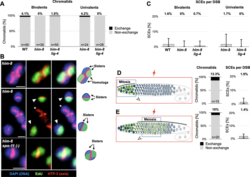
Sexual reproduction shuffles the parental genomes to generate new genetic combinations. To achieve that, the genome is subjected to numerous double-strand breaks, the repair of which involves two crucial decisions: repair pathway and repair template.1 Use of crossover pathways with the homologous chromosome as template exchanges genetic information and directs chromosome segregation. Crossover repair, however, can compromise the integrity of the repair template and is therefore tightly regulated. The extent to which crossover pathways are used during sister-directed repair is unclear because the identical sister chromatids are difficult to distinguish. Nonetheless, indirect assays have led to the suggestion that inter-sister crossovers, or sister chromatid exchanges (SCEs), are quite common.2-11 Here we devised a technique to directly score physiological SCEs in the C. elegans germline using selective sister chromatid labeling with the thymidine analog 5-ethynyl-2'-deoxyuridine (EdU). Surprisingly, we find SCEs to be rare in meiosis, accounting for <2% of repair events. SCEs remain rare even when the homologous chromosome is unavailable, indicating that almost all sister-directed repair is channeled into noncrossover pathways. We identify two mechanisms that limit SCEs. First, SCEs are elevated in the absence of the RecQ helicase BLMHIM-6. Second, the synaptonemal complex-a conserved interface that promotes crossover repair12,13-promotes SCEs when localized between the sisters. Our data suggest that crossover pathways in C. elegans are only used to generate the single necessary link between the homologous chromosomes. Noncrossover pathways repair almost all other breaks, regardless of the repair template.

Keywords: BLM; C. elegans; DNA repair; EdU; RecQ helicase; SCE; crossover; meiosis; synaptonemal complex.


Conflict of interest statement

Declaration of interests The authors declare no competing interests.

Figures

Figure 1:
Figure 1:. A new method to visualize meiotic SCEs using selective labeling with EdU
A. Overview of pulse/chase experiment. Nuclei undergoing DNA replication in the mitotic zone of the gonad incorporate EdU (green). After removal of EdU, the labeled nuclei continue dividing asynchronously until meiotic entry (crescent-shaped nuclei). The labeled nuclei progress through meiosis as they move through the gonad, and are remodeled in diakinesis into condensed structures that allow visualization of individualized sisters. Bottom, an image of a wild-type C. elegans gonad labeled with EdU and chased for 28 hours at 25°C. A labeled nucleus in diakinesis is marked with a dashed box. Note the asynchronous distribution of the labeled nuclei. See Figure S1A for further details. Blue, DNA (DAPI); Green, EdU. B. Schematic illustrating single sister EdU labeling. Chromosomes undergo mitotic DNA replication in the presence of EdU (green), resulting in one EdU-containing strand in each sister. Following pre-meiotic DNA replication without EdU, labeling is reduced to one strand of one sister per homolog pair. In meiotic prophase, the meiotic chromosome axis (pink) appears at the interface between the homologs. When visualized as a condensed bivalent in diakinesis, sister chromatids are clearly resolvable, with the axis now delineating the interface between the sisters. Inset, a confocal image of bivalents harboring or lacking an exchange (left and right, respectively) with interpretative diagrams to the right. The nucleus harboring the exchange bivalent underwent an extra round of DNA replication without EdU before entering meiosis, resulting in labeling of only one of the four sisters. See Figure S1B for a related illustration. Blue, DNA (DAPI); red, axis (anti-HTP-3 antibodies); green, EdU. Scale bars = 1μm. C. EdU incorporation does not cause measurable DNA damage or affect bivalent formation. Analysis of bivalent formation in wild-type and him-8 spo-11(−) worms, which lack meiotic DSBs. (him-8 is present in this background due to genetic linkage with spo-11, but this does not impact the result.) DSB formation in him-8 spo-11(−) animals would result in chiasmata and bivalent formation; no bivalents were observed among the EdU labeled chromosomes. The presence of univalents in wild-type animals would have indicated failure to form chiasmata; no such incidence were observed in the presence or absence of EdU. N values indicate the number of chromosomes counted per genotype. See Figure S2B–C for further analysis.
Figure 2:
Figure 2:. SCEs are rare outcome of meiotic DSB repair
A. Exchange chromatids are rare, regardless of the availability of the homolog. Exchange and non-exchange chromatids on bivalent and univalent chromosomes in wild-type worms, in worms deficient for X chromosome pairing (him-8) and in worms defective for both X chromosome pairing and non-homologous end-joining (him-8; lig-4). N values indicate the number of chromatids counted for each genotype. Results were not significant for all pairwise comparisons with wild-type chromosomes (p>.05, Pearson’s chi-squared test). See also Table S1. B. Representative cytology of bivalents and univalents. Top, a single sister of each homolog pair from him-8 animals labeled with EdU, with no exchanges in either of the long arms. Middle, two unpaired univalent X chromosomes (arrowheads) in him-8 animals, each with a single sister chromatid labeled with EdU, and no exchanges. An autosome bivalent (asterisk) is not labeled due to the different replication time of the X chromosome and the autosomes [62]. Bottom, univalent chromosomes in irradiated him-8 spo-11(−) animals with a single sister chromatid labeled with EdU, and an SCE. Note the EdU signal crossing the axis. Red, axis (anti-HTP-3 antibodies); green, EdU, Blue, DNA (DAPI). Interpretive diagrams are shown on the right. Scale bars = 1μm. C. SCEs are a rare outcome of meiotic DSB repair. Rates of SCE formation per DSB calculated using an estimate of 5 DSBs per homolog pair (2.5 per chromosome). Numbers represent the average rate a DSB resulted in an SCE. Error bars represent 95% exact binomial confidence intervals. D. Direct quantification reveals that SCEs are a rare outcome of exogenous DSBs. Left, cartoon of irradiation experiment. him-8 spo-11(−) worms that are deficient for endogenous DSBs are labeled with EdU and exposed to 5000 rads X-ray irradiation (an average of 7 DSBs per chromosome). While the whole worms were subject to irradiation (red box), the nuclei in the mitotic zone (D) and meiotic prophase (E) reached diakinesis at time of dissection (indicated by blue squares). Middle, observed exchange chromatids. N values indicate the number of chromatids counted for each genotype. Right, rate of SCEs as a fraction of total DSB repair events. Error bars represent 95% exact binomial confidence intervals. See also Table S1.
Figure 3:
Figure 3:. SCEs are elevated in worms lacking the RecQ helicase BLMHIM−6
A. Multiple SCEs are visible in an immunofluorescence image of a bivalent from a him-6 worm. Note that each sister pair underwent an SCE. Red, axis (anti-HTP-3 antibodies); green, EdU, Blue, DNA (DAPI). Interpretive diagram shown to the right. Scale bar = 1μm. B. Elevated levels of meiotic SCEs in him-6 worms. Frequency of observed exchange chromatids in wild-type, him-6 and him-6 spo-11 worms. Note the much lower level of exchanges in him-6 spo-11 versus him-6 worms, indicating that most exchange events in him-6 animals result from meiotic DSBs. Exchange chromosomes in him-6 worms occured at a similar rate in both bivalents and univalents (22/46 and 4/14, respectively). Univalents are formed due to the failure of some inter-homolog crossover precursors to mature to chiasmata [43]. The pairwise comparisons between wild type and him-6 and between him-6 and him-6 spo-11 were significant (p<0.0001 and p<.00001, respectively; Pearson’s Chi-square test). N values indicate the number of chromatids counted. See also Table S1. C. Mostly non-exchange univalents in a representative image of a him-6 spo-11 diakinesis nucleus. Arrowhead indicates the single exchange chromosome. The two X chromosomes (asteriks) are unlabeled due to their late replication. Red, axis (anti-HTP-3 antibodies); green, EdU, Blue, DNA (DAPI). Scale bar = 1μm. D. Premeiotic and meiotic SCE formation in EdU labeled chromatids would result in indistinguishable diakinesis bivalents. Schematics of chromatids undergoing DNA replication with EdU, followed by pre-meiotic DNA replication without EdU, and undergoing SCEs (red asterisks) in meiosis (top) or during premeiotic DNA replication (bottom). Premeiotic SCEs could also occur at times other than premeiotic DNA replication (not shown). See also Figure S4.
Figure 4:
Figure 4:. The Synaptonemal Complex promotes SCEs when it localizes between sister chromatids
A. Many exchange chromatids in an immunofluorescence image of a diakinesis nuclei from a rec-8 animal. Purple, DNA (DAPI), green, EdU. Insets are explained in the interpretive diagrams above and below (boxed regions). Chromatids with crossovers are indicated by arrows. As observed previously [56,57,61], rec-8 mutants affect chromosome morphology in diakinesis, and precluded the use of an axis marker to highlight SCEs. Scale bar = 1μm. B. Univalent chromosomes undergoing multiple SCEs in immunofluorescence images of syp-1K42E worms grown at 25°C. Red, axis (anti-HTP-3 antibodies); green, EdU, Blue, DNA (DAPI). Insets are explained in the interpretive diagrams above and below (boxed regions). Top, diakinesis nucleus containing a univalent with two SCEs. Bottom, a diakinesis nucleus highlighting a univalent with no exchanges (asterisk) and a univalent with a single SCE (arrowhead). See also Figure S3. Scale bar = 1μm. C. Univalent chromosomes undergoing an SCE in immunofluorescence images of syp-3(me42) worms. Red, axis (anti-HTP-3 antibodies); green, EdU, Blue, chromatin (DAPI). Interpretive diagram is shown above. The univalent on the left shows no SCEs, while the univalent on the right harbors an SCE (arrowhead). See also Figure S3. Scale bar = 1μm. D. Elevated frequency of exchange univalents in syp-1K42E, syp-3(me42) and rec-8 worms. The unpaired X univalents in him-8 animals (from Figure 2A) are shown as controls. The percentages above the bars indicate the total number of exchange chromatids (p<0.01, for both pairwise comparisons to him-8, Pearson’s chi-square test; single and double SCEs were pooled together for statistical analysis). N values indicate the number of chromatids counted. See also Table S1. E. A diagram illustrating Synaptonemal Complex localization between sisters in syp-1K42E, syp-3(me42) and rec-8 mutants, thereby promoting SCEs in an analogous way to the promotion of inter-homolog crossovers in wild-type animals. See also Figure S4.

Comment in

Similar articles

Cited by

References

    1. Zickler D, and Kleckner N (2015). Recombination, pairing, and synapsis of homologs during meiosis. Cold Spring Harb. Perspect. Biol. 7, 1–28. - PMC - PubMed
    1. Haber JE, Thorburn PC, and Rogers D (1984). Meiotic and mitotic behavior of dicentric chromosomes in Saccharomyces cerevisiae. Genetics 106, 185–205. - PMC - PubMed
    1. Kadyk LC, and Hartwell LH (1992). Sister chromatids are preferred over homologs as substrates for recombinational repair in Saccharomyces cerevisiae. Genetics 132, 387–402. - PMC - PubMed
    1. Schwacha A, and Kleckner N (1994). Identification of joint molecules that form frequently between homologs but rarely between sister chromatids during yeast meiosis. Cell 76, 51–63. - PubMed
    1. McKim KS, Green-Marroquin BL, Sekelsky JJ, Chin G, Steinberg C, Khodosh R, and Hawley RS (1998). Meiotic synapsis in the absence of recombination. Science 279, 876–878. - PubMed

Publication types

Substances

LinkOut - more resources