. 2021 Sep;32(17-18):949-958.
doi: 10.1089/hum.2020.276. Epub 2021 May 6.

Preclinical Optimization and Safety Studies of a New Lentiviral Gene Therapy for p47phox-Deficient Chronic Granulomatous Disease

Affiliations

Preclinical Optimization and Safety Studies of a New Lentiviral Gene Therapy for p47phox-Deficient Chronic Granulomatous Disease

Andrea Schejtman et al. Hum Gene Ther. 2021 Sep.

Abstract

Chronic granulomatous disease (CGD) is an inherited blood disorder of phagocytic cells that renders patients susceptible to infections and inflammation. A recent clinical trial of lentiviral gene therapy for the most frequent form of CGD, X-linked, has demonstrated stable correction over time, with no adverse events related to the gene therapy procedure. We have recently developed a parallel lentiviral vector for p47phox-deficient CGD (p47phoxCGD), the second most common form of this disease. Using this vector, we have observed biochemical correction of CGD in a mouse model of the disease. In preparation for clinical trial approval, we have performed standardized preclinical studies following Good Laboratory Practice (GLP) principles, to assess the safety of the gene therapy procedure. We report no evidence of adverse events, including mutagenesis and tumorigenesis, in human hematopoietic stem cells transduced with the lentiviral vector. Biodistribution studies of transduced human CD34+ cells indicate that the homing properties or engraftment ability of the stem cells is not negatively affected. CD34+ cells derived from a p47phoxCGD patient were subjected to an optimized transduction protocol and transplanted into immunocompromised mice. After the procedure, patient-derived neutrophils resumed their function, suggesting that gene correction was successful. These studies pave the way to a first-in-man clinical trial of lentiviral gene therapy for the treatment of p47phoxCGD.

Keywords: biodistribution; chronic granulomatous disease; genotoxicity; lentiviral gene therapy.


Conflict of interest statement

The authors declare no conflict of interest. A.J.T. is on the Scientific Advisory Board of Orchard Therapeutics and Rocket Pharmaceuticals. H.L.M. is on the Scientific Advisory Board of Orchard Therapeutics and on the Safety Monitoring Board of Rocket Pharmaceuticals. H.B.G. is Chief Executive Officer for Orchard Therapeutics.

Figures

Figure 1.
Figure 1.
IVIM assay determining the risk of insertional mutagenesis. RF of the control samples Mock or RSF91 and the test vector LV.CHIM-p47 (LV. p47) in comparison to data of a meta-analysis for control samples (MA-Mock, MA-RSF91, MA-Lv-SF). For clarity, the historical data are separated from the experimental ones with a vertical dotted line. The data points below the LOD (plates with no wells above the MTT-threshold) were manually inserted into the graph (due to the logarithmic scale of the y-axis). Above the graph, the ratio of positive (left number) and negative plates (right number) according to the MTT-assay are shown. Differences in the incidence of positive and negative assays relative to MA-Mock or MA-RSF91 were analyzed by Fisher's exact test with BenjaminiHochberg correction (***p < 0.001; NS, not significant). Bars indicate the mean RF. IVIM, in vitro immortalization; LOD, limit of detection; MTT, 3-(4,5-dimethylthiazol-2-yl)-2,5-diphenyltetrazolium bromide (MTT) reduction assay; RF, replating frequency. Color images are available online.
Figure 2.
Figure 2.
Biochemical correction of p47phox−/− mice by lentiviral gene therapy. p47phox−/− Lin cells were transduced with the LV.CHIM-p47 vector at a MOI of 50 (LV.p47low) or 300 (LV.p47high) and transplanted into lethally irradiated p47phox−/−-recipent mice (n = 8). As control we used p47phox−/− mice transplanted with untransduced p47phox−/− (Mock; n = 8) or wild-type (HCT; n = 8) Lin cells and untreated p47phox−/− (KO; n = 4) or C57BL/6 (WT; n = 6) mice. Mice were analyzed up to 6 months after transplantation. (a) VCN and percentage of functional (% of DHR positive) granulocytes in lentivirally transduced p47phox−/− Lin cells upon granulocytic differentiation in liquid cultures (left panel). CFUs and VCN in pooled colonies (right panel). (b) DHR over time in peripheral blood granulocytes of gene therapy-treated mice (LV.p47low and LV.p47high) and of mice in the HCT or Mock groups. (c) VCN in different hematopoietic organs of mice transplanted with LV.p47low (black) or LV.p47 high (red) transduced cells. Data are presented as median and range; Mann–Whitney test, **p < 0.01. (d) Representative DHR plot in granulocytes from the bone marrow of LV.p47 (low and high), Mock, and HCT groups (upper panel). The lower panel shows the correlation between percentage of DHR-positive granulocytes found in the bone marrow of gene therapy-treated p47phox−/− mice and vector copy number (1.5< VCN <7). R2 = 0.66, squared Perason's correlation coefficient, p = 0.0004. (e) p47phox expression in B cells (B220+), T cells (CD3+), monocytes (CD11b+/Gr1low), granulocytes (CD11b+/Gr1 high) in the bone marrow of gene therapy-treated mice (n = 15). Data are mean ± SD; two-way ANOVA with Tukey's multiple comparison, ****p < 0.001. ANOVA, analysis of variance; DHR, dihydrorhodamine; HCT, hematopoietic cell transplantation group; KO, knock out group; MOI, multiplicity of infection; SD, standard deviation; VCN, average vector copy number per cell. Color images are available online.
Figure 3.
Figure 3.
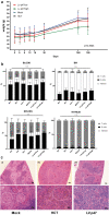
In vivo genotoxicity: body weight, hematological and histopathological analysis of transplanted mice. (a) Monitoring of body weight (g) over time in mice from the LV. p47 low (n = 8), LV. p47 high (n = 7), Mock (n = 7), and HCT (n = 7) groups. Simple linear regression analysis showing no difference between the slopes (p = 0.3923). (b) Percentage of T cells (CD3+), B cells (B220+), and myeloid cells (CD11b+) in PB, BM (KO, n = 4; WT, n = 6; Mock, n = 7; HCT, n = 7; LV.p47low, n = 8; LV.p47high, n = 7), and spleen or thymus (KO, n = 4; WT, n = 3 or n = 6; Mock, n = 7; HCT, n = 6; LV.p47low, n = 8; LV.p47high, n = 6 or n = 5). One hundred percent is given by the total percentage of B, T, and myeloid cells. Data are mean ± SD; two-way ANOVA followed by Tukey's multiple comparison, ns, not significant. (c) Hematoxylin and eosin staining of spleen sections from representative animals belonging to the Mock (“i”, “iv”), HCT (“ii”, “v”), and LV.p47 (“iii”, “vi”) groups at low (upper panels) and high (lower panels) magnification. Areas labeled as “2” show normal red pulp of the spleen, whereas areas labeled as “1” indicate macrophage infiltration. BM, bone marrow. Color images are available online.
Figure 4.
Figure 4.
Vector performance on CD34+ cells from a healthy donor and from a p47phoxCGD patient. (a) CD34+ cells from six healthy donors (each visualized by a different symbol) were transduced with two separate batches of the clinical grade LV.CHIM-p47 vector (p47CGD 18-2-VP-27 and p47CGD 19-I-VM-08; titers of 6.3 × 109 IG/mL and 5.9 × 109 IG/mL, respectively) at different MOI in the presence of 1 mg/mL of LentiBOOST and 4 μg/mL Protamine Sulfate. The x axis shows the range of MOI used and the y axis the VCN. Data are presented as median and range; one-way ANOVA with Tukey's multiple comparison, ns, not significant. (b) p47phoxCGD cells were transduced with the clinical vector (p47CGD 18-2-VP-27) at MOI 130 in the presence of 1 mg/mL of LentiBOOST and 4 μg/mL of Protamine Sulfate, resulting in VCN of 1.84 (shown in brackets). DHR activity (left panel) and p47phox expression (right panel) were assessed by flow cytometry 14 days after culturing CD34+ cells into myeloid differentiation medium (with 50 ng/mL of human GCSF). (c) Transduced p47phoxCGD cells (LV. p47) were transplanted into NSG mice (n = 4) along with nontransduced p47phoxCGD cells (p47CGD; n = 3) and normal donor CD34+ cells (HD; n = 3). Human cell engraftment (%CD45+ cells) was calculated in bone marrow 16 weeks after transplantation. Data are mean ± SD; one-way ANOVA with Tukey's multiple comparison, ns, not significant. (d) DHR activity in CD34+-derived myeloid cells (out of FSC high/CD45+ cells) from pools of LV.p47, p47CGD, and HD mice. CGD, chronic granulomatous disease; FSC, forward scatter; GCSF, granulocyte colony-stimulating factor; HD, healthy donor; NSG, immunodeficient non-obese diabetic (NOD)-SCID Il2rg−/−. Color images are available online.

Similar articles

Cited by

References

    1. Arnold DE, Heimall JR. A review of chronic granulomatous disease. Adv Ther 2017;34:2543–2557. - PMC - PubMed
    1. Rider NL, Jameson MB, Creech CB. Chronic granulomatous disease: epidemiology, pathophysiology, and genetic basis of disease. J Pediatric Infect Dis Soc 2018;7(suppl_1):S2–S5. - PMC - PubMed
    1. Koker MY, Camcioglu Y, van Leeuwen K, et al. . Clinical, functional, and genetic characterization of chronic granulomatous disease in 89 Turkish patients. J Allergy Clin Immunol 2013;132:1156–1163.e5. - PubMed
    1. Tajik S, Badalzadeh M, Fazlollahi MR, et al. . Genetic and molecular findings of 38 Iranian patients with chronic granulomatous disease caused by p47-phox defect. Scand J Immunol 2019;90:e12767. - PubMed
    1. Kutukculer N, Aykut A, Karaca NE, et al. . Chronic granulamatous disease: two decades of experience from a paediatric immunology unit in a country with high rate of consangineous marriages. Scand J Immunol 2019;89:e12737. - PubMed

Publication types

LinkOut - more resources