. 2021 Mar 19;21(1):136.
doi: 10.1186/s12903-021-01502-6.

Tooth mousse containing casein phosphopeptide-amorphous calcium phosphate prevents biofilm formation of Streptococcus mutans

Affiliations

Tooth mousse containing casein phosphopeptide-amorphous calcium phosphate prevents biofilm formation of Streptococcus mutans

Ronit Vogt Sionov et al. BMC Oral Health. .

Abstract

Background: Streptococcus mutans is a common cariogenic bacterium in the oral cavity involved in plaque formation. Casein phosphopeptide-amorphous calcium phosphate (CPP-ACP) has been introduced into tooth mousse to encourage remineralization of dental enamel. The aim of this research was to study the effect of tooth mousse containing CPP-ACP (GC Tooth Mousse®) or CPP-ACP with 0.2% fluoride (CPP-ACPF; GC Tooth Mousse Plus®; GCP) on S. mutans planktonic growth and biofilm formation.

Methods: S. mutans was cultivated in the presence of different dilutions of the tooth mousse containing CPP-ACP or CPP-ACPF, and the planktonic growth was determined by ATP viability assay and counting colony-forming units (CFUs). The resulting biofilms were examined by crystal violet staining, MTT metabolic assay, confocal laser scanning microscopy (CLSM), and scanning electron microscope (SEM).

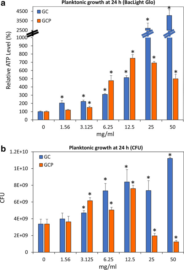
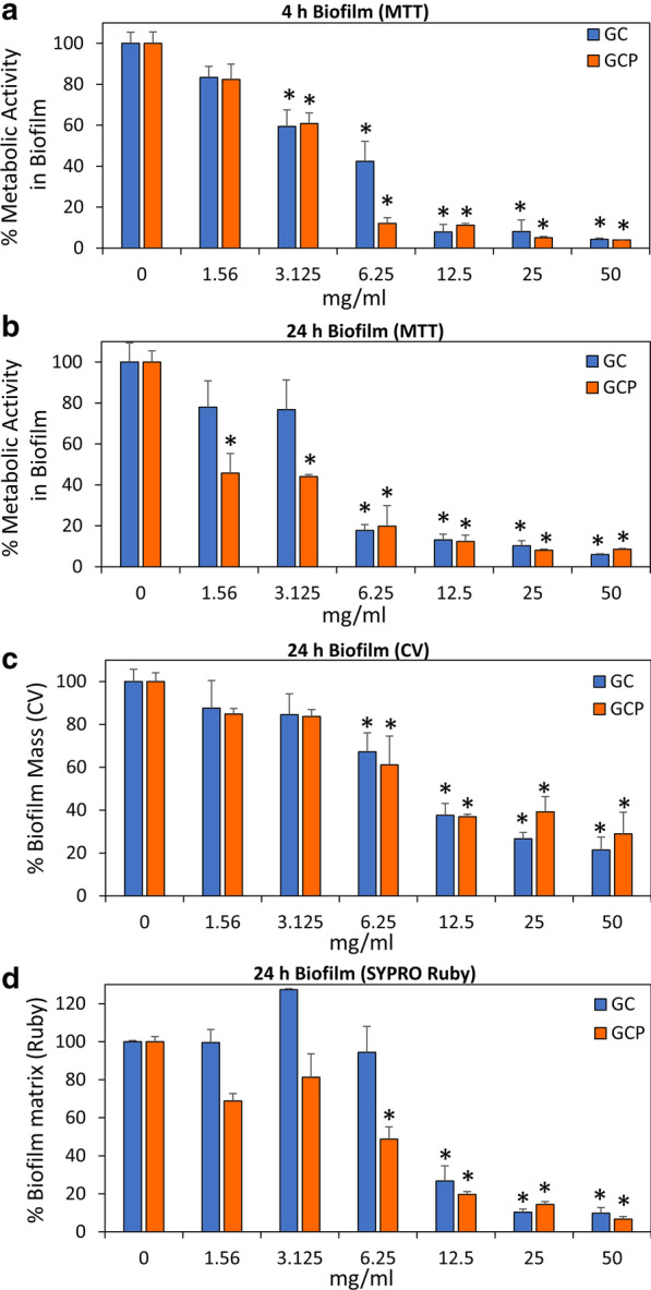
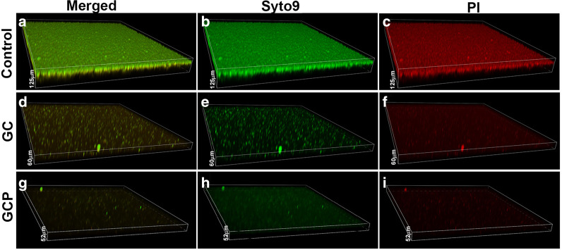
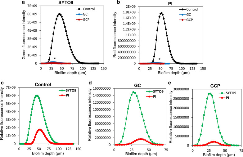
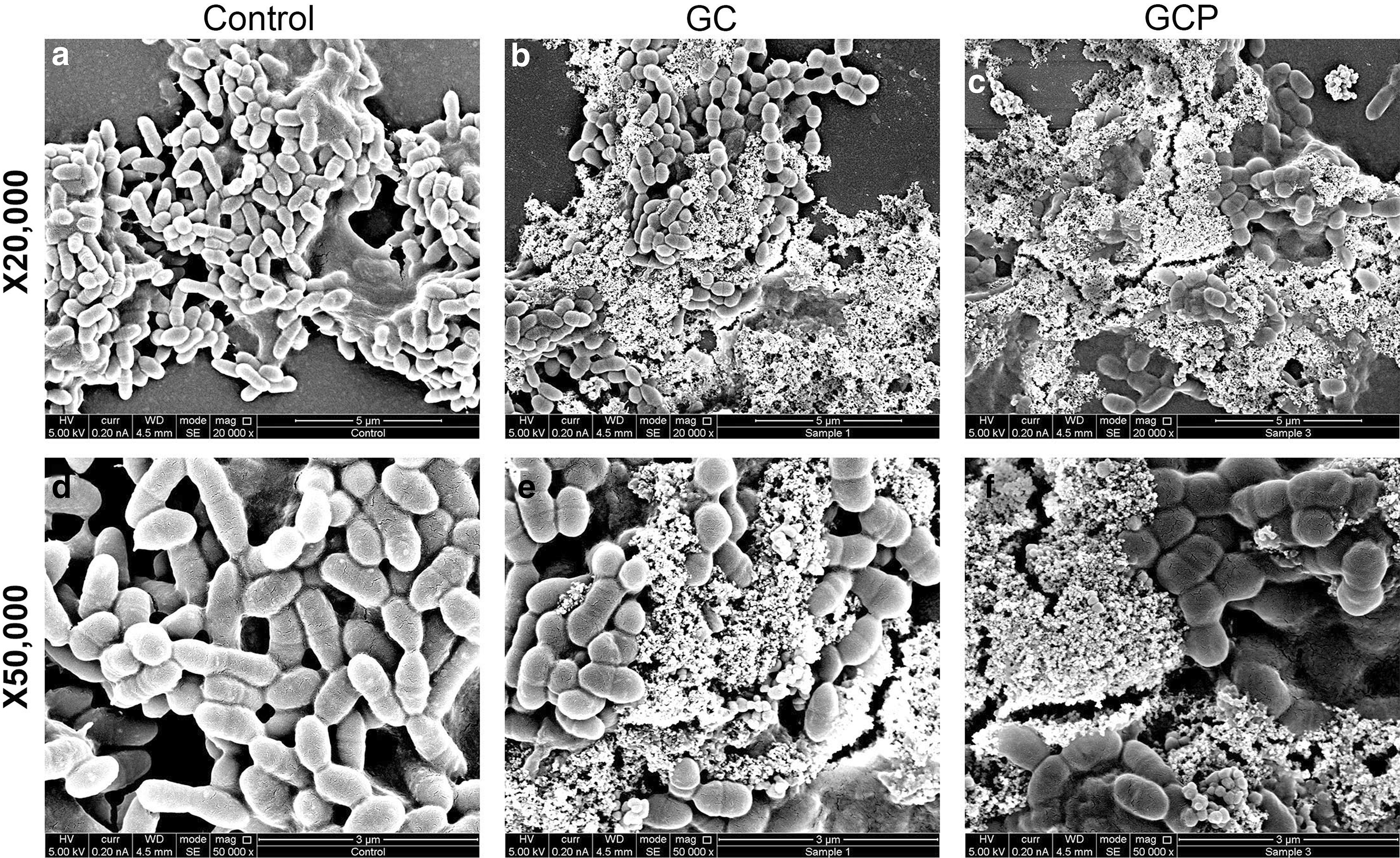
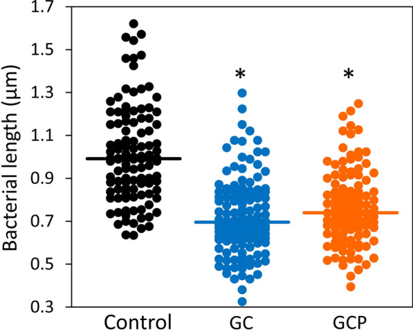
Results: The CPP-ACP tooth mousse (GC) at a dilution of 5-50 mg/ml (0.5-5%) did not inhibit planktonic growth, and even increased the ATP content and the number of viable bacteria after a 24 h incubation. The same was observed for the CPP-ACPF tooth mousse (GCP), except for the higher concentrations (25 and 50 mg/ml) that led to a drop in the bacterial count. Importantly, both compounds significantly decreased S. mutans biofilm formation at dilutions as low as 1.5-3 mg/ml. 12.5 mg/ml GC and 6.25 mg/ml GCP inhibited biofilm formation by 90% after 4 h. After 24 h, the MBIC90 was 6.25 mg/ml for both. CLSM images confirmed the strong inhibitory effect GC and GCP had on biofilm formation when using 5 mg/ml tooth mousse. SEM images of those bacteria that managed to form biofilm in the presence of 5 mg/ml tooth mousse, showed alterations in the bacterial morphology, where the streptococci appear 25-30% shorter on the average than the control bacteria.

Conclusion: Our data show that the tooth mousse containing CPP-ACP reduces biofilm formation of the cariogenic bacterium S. mutans without killing the bacteria. The use of natural substances which inhibit biofilm development without killing the bacteria, has therapeutic benefits, especially in orthodontic pediatric patients.

Keywords: Casein phosphopeptide-amorphous calcium phosphate (CPP-ACP); Dental caries; GC Tooth Mousse®; Oral biofilm; Streptococcus mutans.


Conflict of interest statement

The authors declare no conflict of interest.

Figures

Fig. 1
Fig. 1
The effect of GC and GCP on planktonic growth of S. mutans. a The ATP content of S. mutans that have grown in the presence of various concentrations of suspended GC or GCP tooth mousse for 24 h. n = 3. b The CFU of S. mutans that have grown in the presence of various concentrations of suspended GC or GCP tooth mousse for 24 h. n = 3. * p < 0.05 in comparison to untreated bacteria
Fig. 2
Fig. 2
Anti-biofilm effect of GC and GCP on S. mutans. a Metabolic activity in S. mutans biofilms formed for 4 h in the presence of various concentrations of suspended GC or GCP tooth mousse as determined by the MTT assay. n = 3. b Metabolic activity in S. mutans biofilms formed for 24 h in the presence of various concentrations of suspended GC or GCP tooth mousse as determined by the MTT assay. n = 3. c Crystal violet staining of S. mutans biofilms formed for 24 h in the presence of various concentrations of suspended GC or GCP tooth mousse. n = 3. d FilmTracer™ SYPRO® Ruby biofilm matrix staining of S. mutans biofilms formed for 24 h in the presence of various concentrations of suspended GC or GCP tooth mousse. n = 3. * p < 0.05 in comparison to untreated bacteria
Fig. 3
Fig. 3
CLSM images of SYTO 9/PI-stained biofilms. Biofilms formed in the absence (a–c) or presence of 5 mg/ml GC (d–f) or GCP (g–i) for 24 h were washed in PBS and stained with SYTO 9 (green fluorescence) and PI (red fluorescence). The images are three-dimensional reconstructions of all layers captured using the NIS Element software. a, d, g are merged images of SYTO 9/PI. b, e, h are SYTO 9 staining. c, f, i are PI staining
Fig. 4
Fig. 4
Quantification of the SYTO 9/PI staining of biofilms formed in the absence or presence of GC or GCP using the NIS Element Software. The average of calculations done on 4 different biofilms of each treatment group is presented. a Comparison of SYTO 9 staining of Control (black graph) versus GC (blue graph) and GCP (reddish brown graph)-treated biofilms. b Comparison of PI staining of Control (black graph) versus GC (blue graph) and GCP (reddish brown graph)-treated biofilms. c–e The comparison of SYTO 9 (green graph) and PI (red graph) staining of each treatment group. c Control biofilms. d GC-treated biofilms. e GCP-treated biofilms
Fig. 5
Fig. 5
The effect of GC and GCP on the morphology of S. mutans. S. mutans was allowed to form biofilms for 24 h in the absence (a, d) or presence of 5 mg/ml GC (b, e) or GCP (c, f) for 24 h, and the morphology visualized by HR-SEM. Two different magnifications are shown
Fig. 6
Fig. 6
GC and GCP caused a reduction in the bacterial length of S. mutans. The length of around 100 bacteria were measured in 5 independent HR-SEM images of each treatment group presented in Fig. 5. * p < 0.05 in comparison to untreated bacteria

Similar articles

Cited by

References

    1. Bowen WH, Burne RA, Wu H, Koo H. Oral biofilms: pathogens, matrix, and polymicrobial interactions in microenvironments. Trends Microbiol. 2018;26:229–242. doi: 10.1016/j.tim.2017.09.008. - DOI - PMC - PubMed
    1. Beikler T, Flemmig TF. Oral biofilm-associated diseases: trends and implications for quality of life, systemic health and expenditures. Periodontol. 2011;2000(55):87–103. doi: 10.1111/j.1600-0757.2010.00360.x. - DOI - PubMed
    1. Chen X, Daliri EB, Kim N, Kim JR, Yoo D, Oh DH. Microbial etiology and prevention of dental caries: exploiting natural products to inhibit cariogenic biofilms. Pathogens. 2020;9:569. doi: 10.3390/pathogens9070569. - DOI - PMC - PubMed
    1. Lemos JA, Palmer SR, Zeng L, Wen ZT, Kajfasz JK, Freires IA, Abranches J, Brady LJ. The biology of Streptococcus mutans. Microbiol Spectr. 2019;7(10):1128. - PMC - PubMed
    1. Matsumoto-Nakano M. Role of Streptococcus mutans surface proteins for biofilm formation. Jpn Dent Sci Rev. 2018;54:22–29. doi: 10.1016/j.jdsr.2017.08.002. - DOI - PMC - PubMed

Publication types

Substances

LinkOut - more resources