. 2021 Mar 19;40(1):107.
doi: 10.1186/s13046-021-01906-w.

Exosomal miR-106b-5p derived from melanoma cell promotes primary melanocytes epithelial-mesenchymal transition through targeting EphA4

Affiliations

Exosomal miR-106b-5p derived from melanoma cell promotes primary melanocytes epithelial-mesenchymal transition through targeting EphA4

Wenkang Luan et al. J Exp Clin Cancer Res. .

Abstract

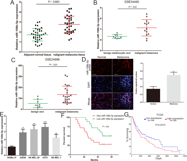
Background: Cancer-secreted exosomal miRNAs regulates the biological processes of many tumours. The serum level of exosomal miR-106b-5p is significantly increased in melanoma patients. However, the role and molecular mechanisms of exosomal miR-106b-5p in melanoma remains unclear.

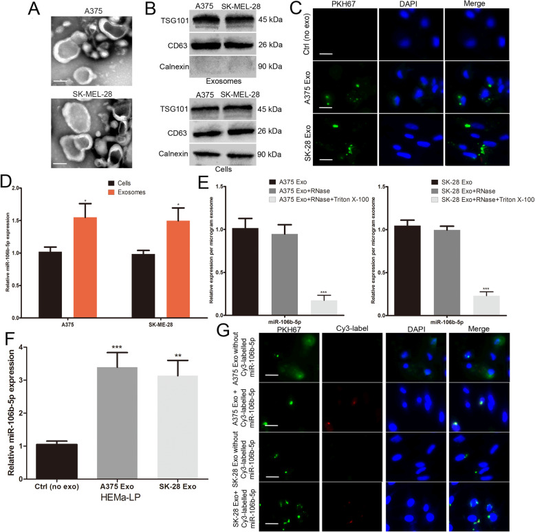
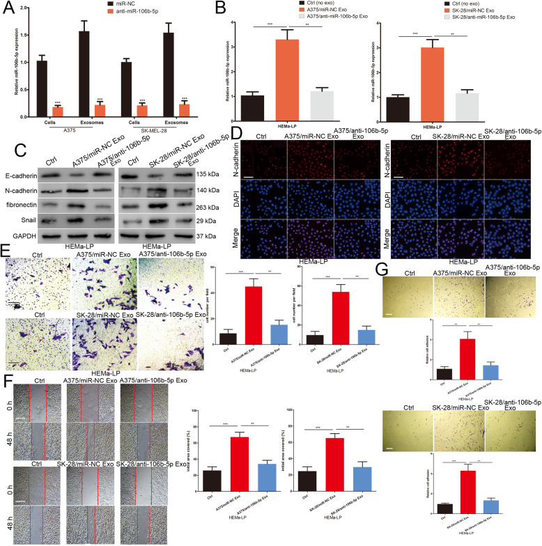
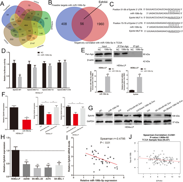
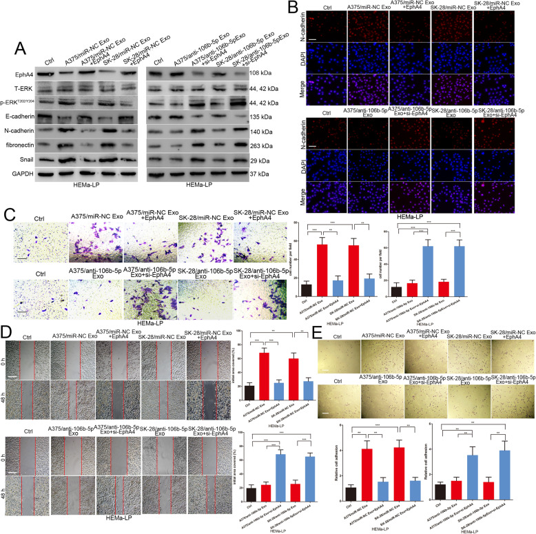
Methods: Quantitative real-time polymerase chain reaction (qRT-PCR) was used to detect the expression of miR-106b-5p and EphA4 in melanoma tissues. Transmission electron microscopy (TEM) and western blotting were used to identify exosome. QRT-qPCR and Cy3-labelled miR-106b-5p were used to demonstrated the transmission of melanoma cell-secreted exosomal miR-106b-5p. Western blotting, Immunofluorescence, adhesion, transwell and scratch wound assay were used to explore the role of exosomal miR-106b-5p in melanocytes. Luciferase reporter assays and RNA-Chromatin Immunoprecipitation (ChIP) assay were used to confirm whether erythropoietin-producing hepatocellular carcinoma receptor A4 (EphA4) was a direct target of miR-106b-5p.

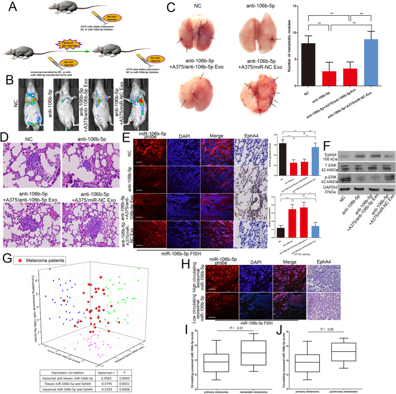
Results: We found that miR-106b-5p levels were increased in melanoma tissue, and high miR-106b-5p expression is an independent risk factor for the overall survival of patients with melanoma. miR-106b-5p is enriched in melanoma cell-secreted exosomes and transferred to melanocytes. Exosomal miR-106b-5p promotes the epithelial-to-mesenchymal transition (EMT), migration, invasion and adhesion of melanocytes. Exosomal miR-106b-5p exerted its role by targeting EphA4 to activate the ERK pathway. We demonstrated that exosomal miR-106b-5p promoted melanoma metastasis in vivo through pulmonary metastasis assay.

Conclusions: Thus, melanoma cell-secreted exosomal miR-106b-5p may serve as a diagnostic indicator and potential therapeutic target in melanoma patients.

Keywords: EMT; EphA4; Exosomal; Melanoma; miR-106b-5p.


Conflict of interest statement

The authors declare that they have no competing interests.

Figures

Fig. 1
Fig. 1
miR-106b-5p expression is upregulated in melanoma and an independent risk factor for the survival of patients with melanoma. a The expression of miR-106b-5p was detected in 36 malignant melanoma tissues and adjacent normal tissues. b The miR-106b-5p levels was analyzed by using GEO#GSE34460 dataset. c The miR-106b-5p levels was analyzed through GEO#GSE24996 dataset. d FISH analysis of miR-106b-5p in malignant melanoma tissues and adjacent normal tissues. Scale bar, 100 μm. e The miR-106b-5p expression profile in human melanoma cell lines (A375, A2058, SK-MEL-28, SK-MEL-1) and human epidermal melanocytes (HEMa-LP). f The overall survival curves of melanoma patients with high miR-106b-5p levels and low miR-106b-5p levels. g The prognostic data of melanoma in TCGA by using OncoLnc (http://www.oncolnc.org). Data were expressed as the mean ± SD, *P < 0.05, **P < 0.01, ***P < 0.001
Fig. 2
Fig. 2
miR-106b-5p is enriched in melanoma cell-secreted exosomes and transferred to melanocytes. a Exosomes purified from culture supernatant of A375 and SK-MEL-28 cells were detected by TEM. Scale bar, 50 nm. b Western blots identified the exosomes marker proteins CD63 and TSG101, and Calnexin was used as an internal reference. c Exosomes purified from culture supernatant of A375 and SK-MEL-28 cells were labeled by PKH67. HEMa-LP cells was co-cultured with these exosomes and observed under confocal microscope, non-exosomes group was used as the negative control. Scale bar, 20 μm. d Basic miR-106b-5p levels in melanoma cells and paired exosomes were detected by qRT-PCR. e miR-106b-5p expression in exosomes, untreated or treated with RNase A and/or Triton X-100. f miR-106b-5p levels in HEMa-LP cells pre-treated with non-exosomes or indicated exosomes for 24 h were detected by qRT-PCR. g Exosomes with Cy3-labeled miR-106b-5p were added to HEMa-LP cells, fluorescence signals were detected under confocal microscope. miR-106b-5p without Cy3-label was used as the negative control. Scale bar, 20 μm. Data were expressed as the mean ± SD, *P < 0.05, **P < 0.01, ***P < 0.001
Fig. 3
Fig. 3
Melanoma cell-secreted exosomal miR-106b-5p promotes the EMT of melanocytes. a Cellular and exosomal miR-106b-5p levels in melanoma cells stably transfected with miR-106b-5p inhibitor or NC lentivectors were detected by qRT-PCR. b miR-106b-5p levels in HEMa-LP cells pre-treated with non-exosomes or melanoma exosomes with different treatment factors. c Western blots identified E-cadherin, N-cadherin, fibronectin and Snail protein expression changes in HEMa-LP cells treated with non-exosomes or indicated exosomes, GAPDH was used as a control. d N-cadherin were identified by immunofluorescence assays in HEMa-LP cells treated with non-exosomes or indicated exosomes. Scale bar, 100 μm. e The invasive capacity of HEMa-LP cells treated with non-exosomes or indicated exosomes was assessed by the transwell assay. Scale bar, 100 μm. f Migration capacity of HEMa-LP cells in different treatment groups was monitored by the scratch wound assay. Scale bar, 200 μm. g The ability of HEMa-LP cells adhesion to fibronectin was detected by the adhesion assay. Scale bar, 100 μm. Data were expressed as the mean ± SD, *P < 0.05, **P < 0.01, ***P < 0.001
Fig. 4
Fig. 4
EphA4 is a direct target of miR-106b-5p. a Bioinformatics software (miRNApath, TargetScan, miRDIP, miRanda and miRDB) predict the potential target genes of miR-106b-5p. b Overlapping candidates with genes that are negatively correlated with miR-106b-5p in the TCGA database. c The binding sites of miR-106b-5p on the 3′-UTR of EphA4, and target sequences of EphA4–3′ UTRs were mutated. d Luciferase assay of cells transfected with EphA4–3′UTR-WT or EphA4–3′UTR-MUT reporter together with miR-106b-5p. e Immunoprecipitation of the Ago2/RISC using the Pan-Ago2 antibody in HEMa-LP cells overexpressing miR-106b-5p. IgG was used as a negative control and β-actin was used as an internal control. qRT-qPCR analysis of miR-106b-5p and EphA4 incorporated into RISC in HEMa-LP cells overexpressing miR-106b-5p compared to the levels in the control. U6 and GAPDH was used as an internal control. f RNA levels of EphA4 were detected by qRT-PCR in HEMa-LP cells treated with indicated exosomes or miR-106b-5p mimic. g Western blot analysis identified EphA4 protein expression changes in HEMa-LP cells treated with indicated exosomes or miR-106b-5p mimic. GAPDH was used as a control. h The EphA4 expression profile in human melanoma cell lines (A375, A2058, SK-MEL-28, SK-MEL-1) and human epidermal melanocytes (HEMa-LP). i The correlation of EphA4 mRNA and miR-106b-5p expression in 36 melanoma tissues was negative. j TCGA dataset revealed a significant negative correlation between EphA4 mRNA and miR-106b-5p in melanoma. Data were expressed as the mean ± SD, *P < 0.05, **P < 0.01, ***P < 0.001
Fig. 5
Fig. 5
Exosomal miR-106b-5p promotes the EMT of melanocytes by targeting EphA4 to activate the ERK pathway. a Western blot analysis of EphA4, E-cadherin, N-cadherin, fibronectin and Snail, total ERK and p-ERKT202/Y204 in HEMa-LP treated with indicated exosomes in the presence of EphA4 plasmid or EphA4 siRNA, GAPDH was used as a control. b N-cadherin were identified by immunofluorescence assays in HEMa-LP cells treated with indicated exosomes in the presence of EphA4 plasmid or EphA4 siRNA. Scale bar, 100 μm. c The invasive capacity of HEMa-LP cells treated with indicated exosomes in the presence of EphA4 plasmid or EphA4 siRNA was assessed by the transwell assay. Scale bar, 100 μm. d Migration ability of HEMa-LP cells in different treatment groups was detected by the scratch wound assay. Scale bar, 200 μm. e The ability of HEMa-LP cells in different treatment groups adhesion to fibronectin was detected by the adhesion assay. Scale bar, 100 μm. Data were expressed as the mean ± SD, *P < 0.05, **P < 0.01, ***P < 0.001
Fig. 6
Fig. 6
Exosomal miR-106b-5p promotes melanoma metastasis in vivo. a The schema of the animal experiment. b Representative bioluminescence images of mice after tail vein injection of stably expressing miR-106b-5p inhibitor A375 cells with or without intravenous injection of exosomes secreted by A375 cells. c The excision lung tissues in nude mice and the number of metastatic lung nodules. d Metastatic lung nodules were confirmed by H&E staining. Scale bar, 25 μm. e The expression of miR-106b-5p and EphA4 were detected by FISH and immunohistochemistry of sections from the metastatic lung nodules. Scale bar, 25 μm. f Western blot analysis of EphA4, total ERK and p-ERKT202/Y204 in metastatic lung nodules, GAPDH was used as a control. g Three dimensional scatter plot of circulating exosomal miR-106b-5p expression, tumour miR-106b-5p levels and EphA4 expression in 36 melanoma patients. h The expression of miR-106b-5p and EphA4 were detected by FISH and immunohistochemistry of sections from the malignant melanoma tissues. Scale bar, 25 μm. i The expression of circulating exosomal miR-106b-5p in metastatic and primary melanoma patients j the circulating exosomal miR-106b-5p level in 7 pulmonary metastasis patients and primary melanoma patients. Data were expressed as the mean ± SD, *P < 0.05, **P < 0.01, ***P < 0.001

Similar articles

Cited by

References

    1. Siegel RL, Miller KD, Jemal A. Cancer statistics, 2017. CA Cancer J Clin. 2017;67(1):7–30. doi: 10.3322/caac.21387. - DOI - PubMed
    1. Chen W, Zheng R, Baade PD, Zhang S, Zeng H, Bray F, Jemal A, Yu XQ, He J. Cancer statistics in China, 2015. CA Cancer J Clin. 2016;66(2):115–132. doi: 10.3322/caac.21338. - DOI - PubMed
    1. Little EG, Eide MJ. Update on the current state of melanoma incidence. Dermatol Clin. 2012;30(3):355–361. doi: 10.1016/j.det.2012.04.001. - DOI - PubMed
    1. Haass NK, Schumacher U. Melanoma never says die. Exp Dermatol. 2014;23(7):471–472. doi: 10.1111/exd.12400. - DOI - PubMed
    1. Tsao H, Chin L, Garraway LA, Fisher DE. Melanoma: from mutations to medicine. Genes Dev. 2012;26(11):1131–1155. doi: 10.1101/gad.191999.112. - DOI - PMC - PubMed