. 2021 Mar 19;14(1):46.
doi: 10.1186/s13045-021-01059-5.

Low expression of TRAF3IP2-AS1 promotes progression of NONO-TFE3 translocation renal cell carcinoma by stimulating N6-methyladenosine of PARP1 mRNA and downregulating PTEN

Affiliations

Low expression of TRAF3IP2-AS1 promotes progression of NONO-TFE3 translocation renal cell carcinoma by stimulating N6-methyladenosine of PARP1 mRNA and downregulating PTEN

Lei Yang et al. J Hematol Oncol. .

Erratum in

Abstract

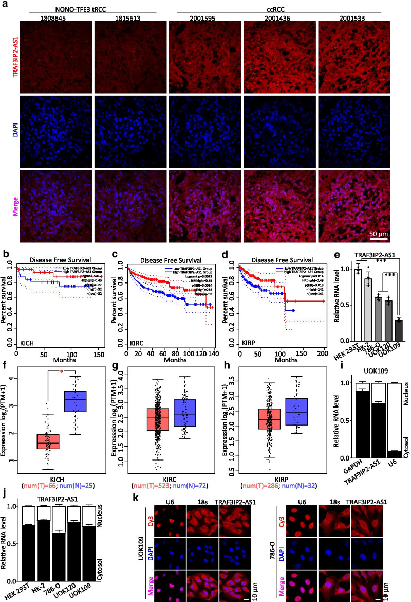
Background: NONO-TFE3 translocation renal cell carcinoma (NONO-TFE3 tRCC) is one subtype of RCCs associated with Xp11.2 translocation/TFE3 gene fusions RCC (Xp11.2 tRCCs). Long non-coding RNA (lncRNA) has attracted great attention in cancer research. The function and mechanisms of TRAF3IP2 antisense RNA 1 (TRAF3IP2-AS1), a natural antisense lncRNA, in NONO-TFE3 tRCC remain poorly understood.

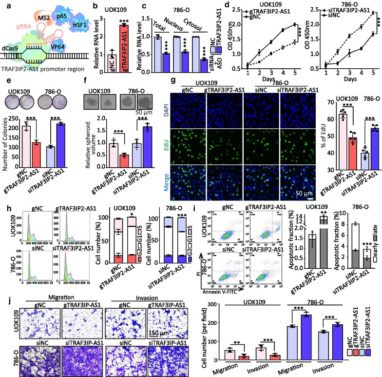
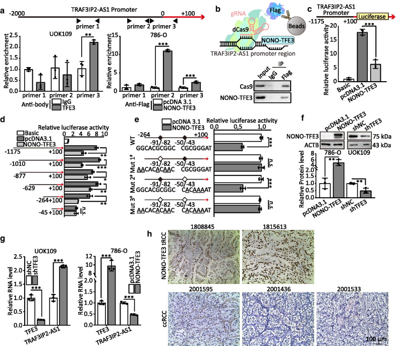
Methods: FISH and qRT-PCR were undertaken to study the expression, localization and clinical significance of TRAF3IP2-AS1 in Xp11.2 tRCC tissues and cells. The functions of TRAF3IP2-AS1 in tRCC were investigated by proliferation analysis, EdU staining, colony and sphere formation assay, Transwell assay and apoptosis analysis. The regulatory mechanisms among TRAF3IP2-AS1, PARP1, PTEN and miR-200a-3p/153-3p/141-3p were investigated by luciferase assay, RNA immunoprecipitation, Western blot and immunohistochemistry.

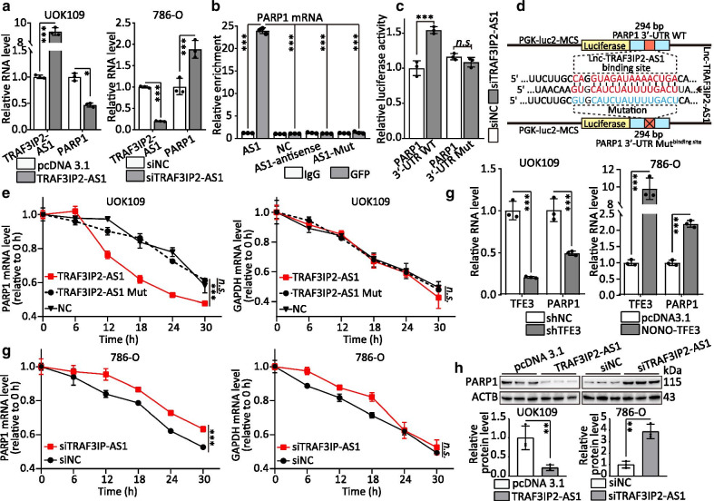
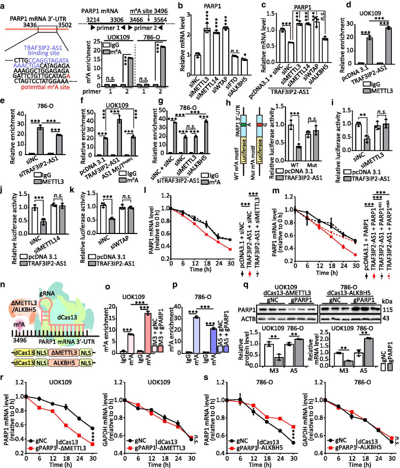
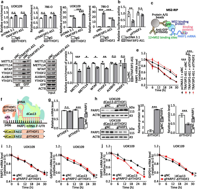
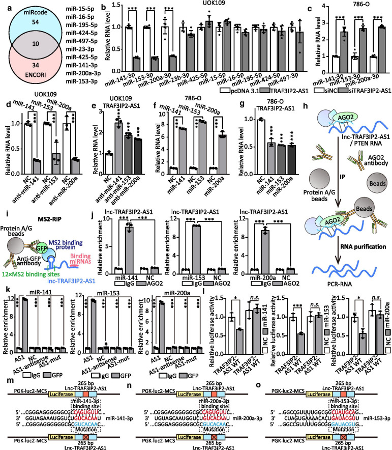
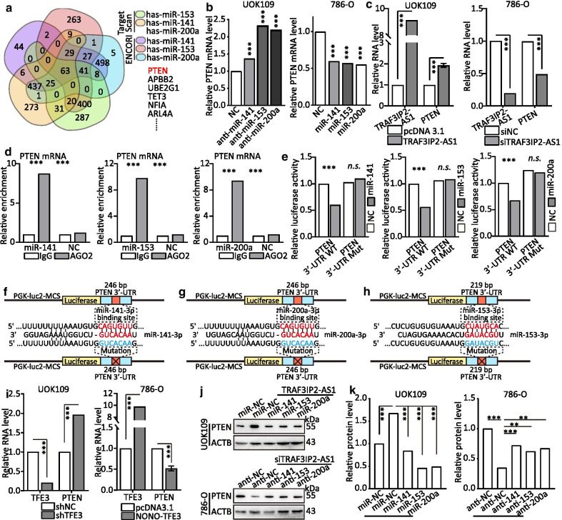
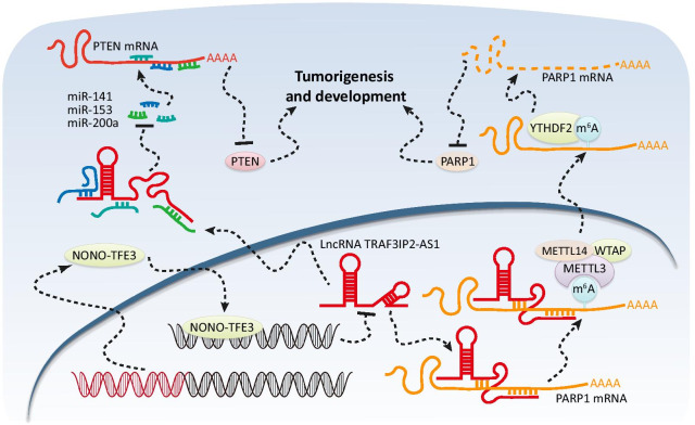
Results: The expression of TRAF3IP2-AS1 was suppressed by NONO-TFE3 fusion in NONO-TFE3 tRCC tissues and cells. Overexpression of TRAF3IP2-AS1 inhibited the proliferation, migration and invasion of UOK109 cells which were derived from cancer tissue of patient with NONO-TFE3 tRCC. Mechanistic studies revealed that TRAF3IP2-AS1 accelerated the decay of PARP1 mRNA by direct binding and recruitment of N6-methyladenosie methyltransferase complex. Meanwhile, TRAF3IP2-AS1 competitively bound to miR-200a-3p/153-3p/141-3p and prevented those from decreasing the level of PTEN.

Conclusions: TRAF3IP2-AS1 functions as a tumor suppressor in NONO-TFE3 tRCC progression and may serve as a novel target for NONO-TFE3 tRCC therapy. TRAF3IP2-AS1 expression has the potential to serve as a novel diagnostic and prognostic biomarker for NONO-TFE3 tRCC detection.

Keywords: M6A modification; NONO-TFE3; PARP1; PTEN; TRAF3IP2-AS1.


Conflict of interest statement

The authors declare that they have no competing interests.

Figures

Fig. 1
Fig. 1
TRAF3IP2-AS1 expression is significantly decreased in NONO-TFE3 tRCC. a The RNA level of TRAF3IP2-AS1 (red) was analyzed by FISH assays in NONO-TFE3 tRCC and ccRCC. DAPI-stained nuclei are blue. b–d Kaplan–Meier analysis revealed the disease-free survival (DFS) in KICH, KIRC and KIRP patients based on the relative TRAF3IP2-AS1 expression. e The RNA level of TRAF3IP2-AS1 was analyzed by qRT-PCR assay in ccRCC cell line (786-O), tRCC cell lines (UOK109 and UOK120) and normal cell lines (HK-2 and HEK293T). f–h Analysis of TRAF3IP2-AS1 in KICH, KIRC and KIRP tissues compared with normal tissues were performed using TCGA data. i–j The subcellular distribution of TRAF3IP2-AS1 was analyzed via qRT-PCR in HEK293T, HK-2, 786-O, UOK120 and UOK109 cells. U6 and GAPDH were used as nuclear and cytoplasmic markers, respectively. k The location of TRAF3IP2-AS1 (red) in UOK109 and 786-O cells was determined by FISH assay. U6 and 18s rRNA were used as positive controls for the nuclear and cytoplasmic fractions, respectively. DAPI-stained nuclei are blue. The data are presented as the mean ± SD, *P < 0.05, ***P < 0.001
Fig. 2
Fig. 2
TRAF3IP2-AS1 deficiency induces development of NONO-TFE3 tRCC. a Schematic illustration of CRISPR/Cas9-based Synergistic Activation Mediator (SAM) system. b The RNA level of TRAF3IP2-AS1 was analyzed by qRT-PCR assay in UOK109 transfected with dCas9 and guide RNA targeting TRAF3IP2-AS1 promoter (gTRAF3IP2-AS1). c The RNA level of TRAF3IP2-AS1 was analyzed by qRT-PCR assay in 786-O transfected with siRNA and antisense oligonucleotides (ASOs). d–f The effects of TRAF3IP2-AS1 overexpression or knockdown on the proliferation of UOK109 and 786-O cells, respectively, were examined by CCK-8 assay (d), colony formation assays (e) and tumor sphere formation (f). g EdU assays were used to detect the proliferation rate of UOK109 and 786-O cells after transfection for 48 h. h Cell cycle was analyzed using flow cytometry after transfection for 48 h. i Cell apoptosis was analyzed via flow cytometry using an Annexin V/PI Kit after transfection for 48 h. j Migration and invasion assays were performed with transfected cells using Transwell inserts. The data are presented as the mean ± SD, *P < 0.05, **P < 0.01, ***P < 0.001
Fig. 3
Fig. 3
NONO-TFE3 inhibits lncRNA TRAF3IP2-AS1 transcription. a ChIP assays showed endogenous NONO-TFE3 binding to the TRAF3IP2-AS1 gene promoter. 786-O cells were transfected with NONO-TFE3-Flag-overexpressing vector for 48 h. The binding of NONO-TFE3 at the TRAF3IP2-AS1 promoter region was detected by a ChIP assay. b Schematic summary of the dCas9-gRNA-guided ChIP (upper); Western blot was performed after dCas9-gRNA-guided ChIP (lower). c–d HEK293T cells were co-transfected with TRAF3IP2-AS1 promoter–luciferase truncations and NONO-TFE3 plasmids, and the luciferase activity was determined using a Dual Luciferase Reporter Assay after 48 h. e Dual luciferase assay of HEK293T cells co-transfected with firefly luciferase constructs containing the wild-type or mutant NONO-TFE3 potential binding sites of TRAF3IP2-AS1 promoter and NONO-TFE3 plasmids were performed. g The protein and mRNA levels of NONO-TFE3 and the TRAF3IP2-AS1 expression levels were detected after transfection with NONO-TFE3 plasmids or shTFE3 for 48 h. h NONO-TFE3 immunohistochemistry was performed in paraffin sections of samples from patients. The data are presented as the mean ± SD, *P < 0.05, **P < 0.01, ***P < 0.001
Fig. 4
Fig. 4
TRAF3IP2-AS1 down-regulates PARP1 mRNA by direct binding. a The RNA levels of TRAF3IP2-AS1 and PARP1 were detected after transfection with TRAF3IP2-AS1 plasmids or siTRAF3IP2-AS1 for 48 h. b Level of PARP1 mRNA detected by qRT-PCR after MS2-RIP for GFP in UOK109 cells. AS1, NC, AS1-antisense and AS1-Mut correspond to TRAF3IP2-AS1, empty vector, TRAF3IP2-AS1-antisense and TRAF3IP2-AS1 with mutation of potential binding site to PARP1 mRNA, respectively. c–d Dual Luciferase Reporter Assay used to detect the relative luciferase activity in HEK293 T cells co-transfected with siTRAF3IP2-AS1 and pmirGLO-PARP1 3′-UTR WT/MUT. e The stability of PARP1 mRNA and GAPDH mRNA in UOK109 cells transfected with TRAF3IP2-AS1 or TRAF3IP2-AS1 contained binding site mutation was measured by qRT-PCR relative to 0 h after blocking new RNA synthesis with α-amanitin and normalized to 18 s rRNA. f The RNA levels of NONO-TFE3 and PARP1 were detected after transfection with NONO-TFE3 plasmids or shTFE3 for 48 h. g The stability of PARP1 mRNA and GAPDH mRNA in 786-O cells transfected with siTRAF3IP2-AS1 was measured by qRT-PCR after treatment with α-amanitin. h The protein level of PARP1 was detected after transfection with TRAF3IP2-AS1 plasmids or siTRAF3IP2-AS1
Fig. 5
Fig. 5
TRAF3IP2-AS1 accelerates the decay of PARP1 mRNA by recruitment of m6A methyltransferase complex. a The location of TRAF3IP2-AS1 binding site and a potential m6A site are indicated. Abundance of PARP1 transcript among mRNA immunoprecipitated with anti-m6A antibody was measured by qRT-PCR and normalized to IgG. b–c The mRNA level of PARP1 was detected by qRT-PCR after transfected with indicated vectors or siRNAs. d–e METTL3 was immunoprecipitated followed by qRT-PCR for assessing the association of the indicated PARP1 mRNA with METTL3 after overexpression or knockdown of TRAF3IP2-AS1. f–g Abundance of PARP1 among mRNA immunoprecipitated with anti-m6A antibody from cells transfected with indicated vectors or siRNA/ASO was measured by qRT-PCR. h PARP1-3′-UTR of the wild-type or containing a m6A consensus sequence mutant (A to G) was fused with a luciferase reporter. Luciferase activity of PARP1-3′-UTR was measured and normalized to Renilla luciferase activity. p–k Luciferase activity of PARP1-3′-UTR was measured after co-transfected with TRAF3IP3-AS1 and siMETTL3/siMEETTL14/siWTAP. l–m The stability of PARP1 mRNA in cells co-transfected with indicated vectors or siRNAs after treatment with α-amanitin. n Schematic illustration of targeted RNA methylation system. o–p Abundance of PARP1 among mRNA immunoprecipitated with anti-m6A antibody from cells transfected with indicated gRNA was measured by qRT-PCR. q The protein and mRNA level of PARP1 were measured by qRT-PCR and Western blot after transfected with indicated gRNA. r–s The stability of PARP1 and GAPDH mRNA in cells transfected with indicated gRNA after treatment with α-amanitin. The data are presented as the mean ± SD, *P < 0.05, **P < 0.01, ***P < 0.001
Fig. 6
Fig. 6
YTHDF2 mediates the decay of PARP1 mRNA to regulate NONO-TFE3 tRCC progression. a YTHDF1/2 was immunoprecipitated followed by qRT-PCR for assessing the association of PARP1 mRNA with YTHDF1/2 after overexpression or knockdown of TRAF3IP2-AS1. b Luciferase activity of PARP1-3′-UTR was measured after co-transfected with TRAF3IP3-AS1 and siYTHDF2. c Schematic illustration of MS2-RIP. d Abundance of m6A relative proteins among MS2-RIP with anti-GFP antibody from cells transfected with indicated plasmid was measured by Western blot. e The stability of PARP1 mRNA in cells co-transfected with indicated vectors or siRNAs after treatment with α-amanitin. f Schematic illustration of targeted RNA methylation system. g–h The mRNA (g) and protein (h) level of PARP1 were measured by qRT-PCR and Western blot after transfected with indicated gRNA. i–j The stability of PARP1 and GAPDH mRNA in cells transfected with indicated gRNA after treatment with α-amanitin. The data are presented as the mean ± SD, *P < 0.05, **P < 0.01, ***P < 0.001
Fig. 7
Fig. 7
TRAF3IP2-AS1 functions as a ceRNA and sponges miRNAs. a Schematic of the selection for the direct downstream target of TRAF3IP2-AS1. b–c The effect of TRAF3IP2-AS1 on multiple miRNAs expression in UOK109 cells was analyzed by qRT-PCR after overexpression or knockdown of TRAF3IP2-AS1. d–f The RNA levels of multiple miRNAs and TRAF3IP2-AS1 were analyzed via qRT-PCR in UOK109 cells after transfected with miRNA inhibitor, respectively. e–g The RNA levels of multiple miRNAs and TRAF3IP2-AS1 were analyzed via qRT-PCR in 786-O cells after overexpression miRNAs, respectively. h–i Model of AGO2-RIP/MS2-RIP assay. j RIP assays were performed using AGO2 antibody in UOK109 cells, and then, the enrichment ofTRAF3IP2-AS1was detected by qRT-PCR. k MS2-RIP-derived RNA was examined by qRT-PCR. The levels of the qRT-PCR products were normalized relative to IgG control. l HEK293T cells were co-transfected with miRNA mimics, respectively, and wild-type or mutant TRAF3IP2-AS1 luciferase reporter vector, and luciferase reporter activity was detected. m–o Schematic illustration of TRAF3IP2-AS1 wild type and mutation luciferase reporter vectors. The data are presented as the mean ± SD, *P < 0.05, **P < 0.01, ***P < 0.001
Fig. 8
Fig. 8
TRAF3IP2-AS1 modulates the expression of PTEN through post-transcriptional regulation of miRNAs. a Schematic of the selection for the direct downstream target of TRAF3IP2-AS1. b–c The effect of multiple miRNAs expression on PTEN mRNA in UOK109 cells was analyzed by qRT-PCR after overexpression or knockdown of miRNAs, respectively. d RIP assays were performed using AGO2 antibody in UOK109 cells, and then, the enrichment of PTEN mRNA was detected by qRT-PCR. e HEK293T cells were co-transfected with miRNA mimics, respectively, and wild-type or mutant PTEN 3′-UTR luciferase reporter vector, and luciferase reporter activity was detected. f–h Schematic illustration of PTEN 3′-UTR wild type and mutation luciferase reporter vectors. i The mRNA levels of NONO-TFE3 and PTEN were detected after transfection with NONO-TFE3 plasmids or shTFE3 for 48 h. j–k The protein level of PTEN was detected after co-transfection with indicated vectors, siRNAs/ASOs or miRNAs inhibitor for 48 h. The data are presented as the mean ± SD, **P < 0.01, ***P < 0.001
Fig. 9
Fig. 9
Schematic diagram for the mechanisms of TRAF3IP2-AS1 functioning as both an mRNA decay accelerator and a miRNA sponge to inhibit progression of NONO-TFE3 tRCC

References

    1. Moch H, Cubilla AL, Humphrey PA, Reuter VE. Ulbright TMJEU: the 2016 WHO classification of tumours of the urinary system and male genital organs—part A: renal, penile, and testicular. Tumours. 2016;70(1):93–105. - PubMed
    1. Argani P, Lui MY, Couturier J, Bouvier R, Fournet JC, Ladanyi M. A novel CLTC-TFE3 gene fusion in pediatric renal adenocarcinoma with t(X;17)(p11.2;q23) Oncogene. 2003;22(34):5374–5378. doi: 10.1038/sj.onc.1206686. - DOI - PubMed
    1. Argani P, Zhong M, Reuter VE, Fallon JT, Epstein JI, Netto GJ, Antonescu CR. TFE3-fusion variant analysis defines specific clinicopathologic associations among Xp11 translocation cancers. Am J Surg Pathol. 2016;40(6):723–737. doi: 10.1097/PAS.0000000000000631. - DOI - PMC - PubMed
    1. Argani P, Antonescu CR, Couturier J, Fournet JC, Sciot R, Debiec-Rychter M, Hutchinson B, Reuter VE, Boccon-Gibod L, Timmons C, et al. PRCC-TFE3 renal carcinomas: morphologic, immunohistochemical, ultrastructural, and molecular analysis of an entity associated with the t(X;1)(p11.2;q21) Am J Surg Pathol. 2002;26(12):1553–1566. doi: 10.1097/00000478-200212000-00003. - DOI - PubMed
    1. Clark J, Lu YJ, Sidhar SK, Parker C, Gill S, Smedley D, Hamoudi R, Linehan WM, Shipley J, Cooper CS. Fusion of splicing factor genes PSF and NonO (p54nrb) to the TFE3 gene in papillary renal cell carcinoma. Oncogene. 1997;15(18):2233–2239. doi: 10.1038/sj.onc.1201394. - DOI - PubMed

Publication types

MeSH terms