Review
. 2022 Jan-Feb;67(1):252-270.
doi: 10.1016/j.survophthal.2021.03.003. Epub 2021 Mar 16.

Artificial intelligence: the unstoppable revolution in ophthalmology

Affiliations
Review

Artificial intelligence: the unstoppable revolution in ophthalmology

David Benet et al. Surv Ophthalmol. 2022 Jan-Feb.

Abstract

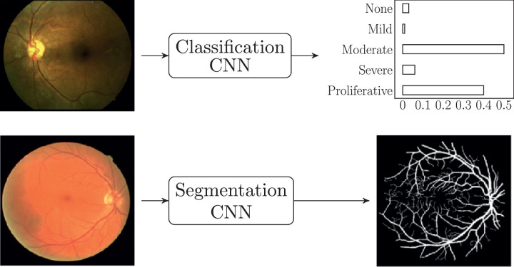
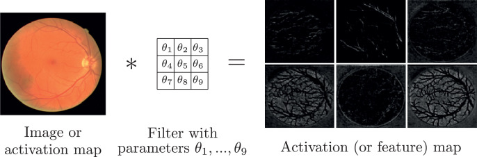
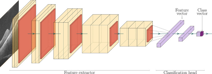
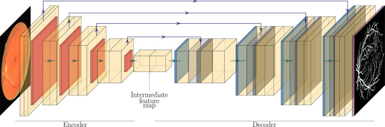
Artificial intelligence (AI) is an unstoppable force that is starting to permeate all aspects of our society as part of the revolution being brought into our lives (and into medicine) by the digital era, and accelerated by the current COVID-19 pandemic. As the population ages and developing countries move forward, AI-based systems may be a key asset in streamlining the screening, staging, and treatment planning of sight-threatening eye conditions, offloading the most tedious tasks from the experts, allowing for a greater population coverage, and bringing the best possible care to every patient. This paper presents a review of the state of the art of AI in the field of ophthalmology, focusing on the strengths and weaknesses of current systems, and defining the vision that will enable us to advance scientifically in this digital era. It starts with a thorough yet accessible introduction to the algorithms underlying all modern AI applications. Then, a critical review of the main AI applications in ophthalmology is presented, including diabetic retinopathy, age-related macular degeneration, retinopathy of prematurity, glaucoma, and other AI-related topics such as image enhancement. The review finishes with a brief discussion on the opportunities and challenges that the future of this field might hold.

Keywords: Age-related macular degeneration; Artificial intelligence; Deep learning; Diabetic retinopathy; Glaucoma; Machine learning; Ophthalmology; Optical coherence tomography; Retina; Retinopathy of prematurity.


Figures

Fig. 1
Fig. 1
Supervised learning.
Fig. 2
Fig. 2
Linear regression model.
Fig. 3
Fig. 3
Logistic regression model.
Fig. 4
Fig. 4
Feed forward neural network for regression with a two-neuron hidden layer.
Fig. 5
Fig. 5
Good fitting (left) compared to overfitting (right).
Fig. 6
Fig. 6
Types of CNNs: classification (above) and segmentation (below). Images from Sayres et al. and Arsalan et al. .
Fig. 7
Fig. 7
Convolution: an image (or an activation map) is convolved with a filter (with learnable parameters θ), to produce an activation map (also called feature map). Images from Arsalan et al. .
Fig. 8
Fig. 8
VGG16 architecture. OCT image from Antony et al. .
Fig. 9
Fig. 9
U-Net architecture. Fundus image and vessel segmentation mask from Arsalan et al. .
Fig. 10
Fig. 10
Class activation maps for a CNN trained on AMD classification. The colored regions are important for the CNN to perform the classification task. Image from Antony et al. .
Fig. 11
Fig. 11
3D CNN for OCT B-scan segmentation. Images from De Fauw et al. .
Fig. 12
Fig. 12
Recurrent neural network.
Fig. 13
Fig. 13
Decision tree for classification.

References

    1. Cheloni R., Gandolfi S.A., Signorelli C., Odone A. Global prevalence of diabetic retinopathy: protocol for a systematic review and meta-analysis. BMJ Open. 2019;9(3) doi: 10.1136/bmjopen-2018-022188. - DOI - PMC - PubMed
    1. Williams R., Karuranga S., Malanda B., et al. 9th edition. Vol. 162. Diabetes Res Clin Pract; 2020. (Global and regional estimates and projections of diabetes-related health expenditure: results from the International Diabetes Federation Diabetes Atlas). - DOI - PubMed
    1. Yang W.H., Zheng B., Wu M.N., et al. An evaluation system of fundus photograph-based intelligent diagnostic technology for diabetic retinopathy and applicability for research. Diabetes Ther. 2019;10(5):1811–1822. doi: 10.1007/s13300-019-0652-0. - DOI - PMC - PubMed
    1. Hartnett M.E. Retinopathy of prematurity: evolving treatment with anti-vascular endothelial growth factor. Am J Ophthalmol. 2020 doi: 10.1016/j.ajo.2020.05.025. Published online. - DOI - PMC - PubMed
    1. Varadarajan A.V., Bavishi P., Ruamviboonsuk P., et al. Predicting optical coherence tomography-derived diabetic macular edema grades from fundus photographs using deep learning. Nat Commun. 2020;11(1):1–8. doi: 10.1038/s41467-019-13922-8. - DOI - PMC - PubMed

Further reading

    1. Brown J.M., Campbell J.P., Beers A., et al. Automated diagnosis of plus disease in retinopathy of prematurity using deep convolutional neural networks. JAMA Ophthalmology. 2018;136:803–810. doi: 10.1001/jamaophthalmol.2018.1934. American Medical Association. - DOI - PMC - PubMed
    1. Brown T.B., Mann B., Ryder N., et al. Language models are few-shot learners. arXiv. 2020;1(May):1–7. https://arxiv.org/abs/2005.14165 Available at: Accessed January 22, 2021.
    1. Burlina P.M., Joshi N., Pekala M., Pacheco K.D., Freund D.E., Bressler N.M. Automated grading of age-related macular degeneration from color fundus images using deep convolutional neural networks. JAMA Ophthalmol. 2017;135(11):1170–1176. doi: 10.1001/jamaophthalmol.2017.3782. - DOI - PMC - PubMed
    1. Cao K., Xu J., Zhao W.Q. Artificial intelligence on diabetic retinopathy diagnosis: an automatic classification method based on grey level co-occurrence matrix and naive Bayesian model. Int J Ophthalmol. 2019;12(7):1158–1162. doi: 10.18240/ijo.2019.07.17. - DOI - PMC - PubMed
    1. Coyner A.S., Swan R., Campbell J.P., et al. Automated fundus image quality assessment in retinopathy of prematurity using deep convolutional neural networks. Ophthalmol Retin. 2019;3(5):444–450. doi: 10.1016/j.oret.2019.01.015. - DOI - PMC - PubMed