

Juvenile Psammomatoid Cemeto-ossifying Fibroma of Mandible: a Diagnostic dilemma
- PMID: 33741572
- PMCID: PMC7986957
- DOI: 10.1136/bcr-2020-240952
Juvenile Psammomatoid Cemeto-ossifying Fibroma of Mandible: a Diagnostic dilemma
Abstract
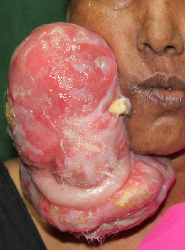
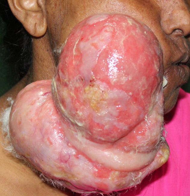
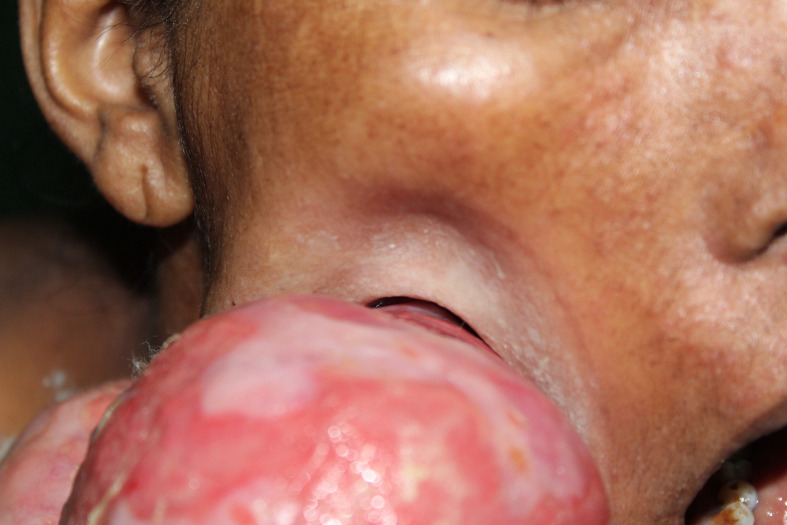
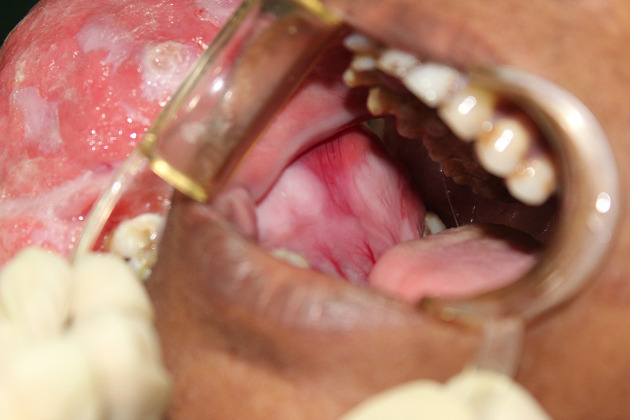
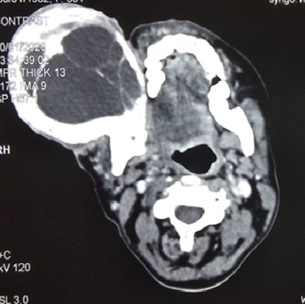
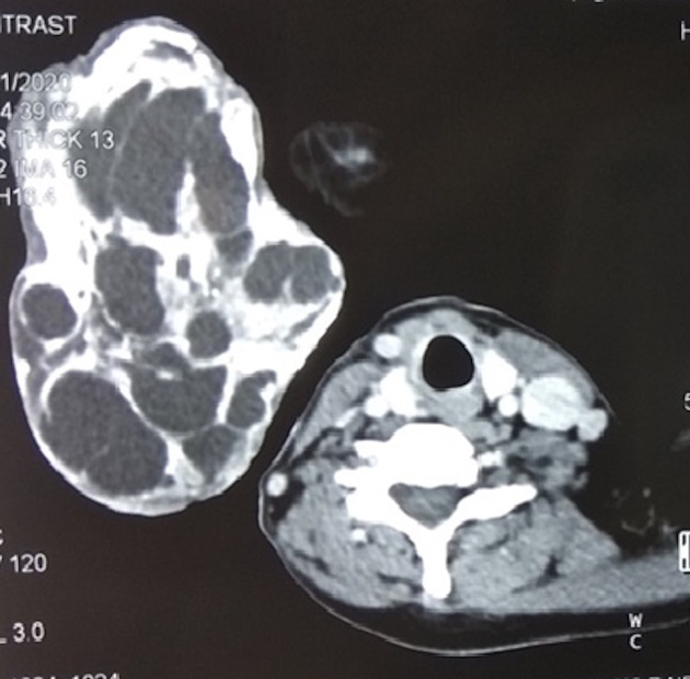
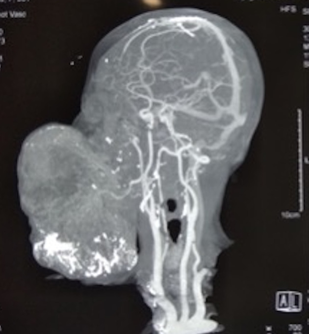
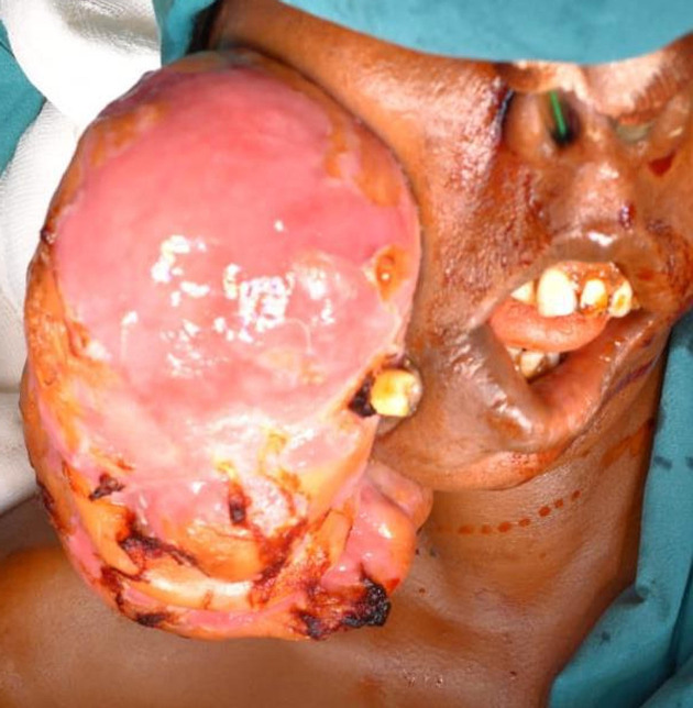
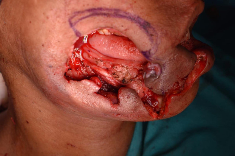
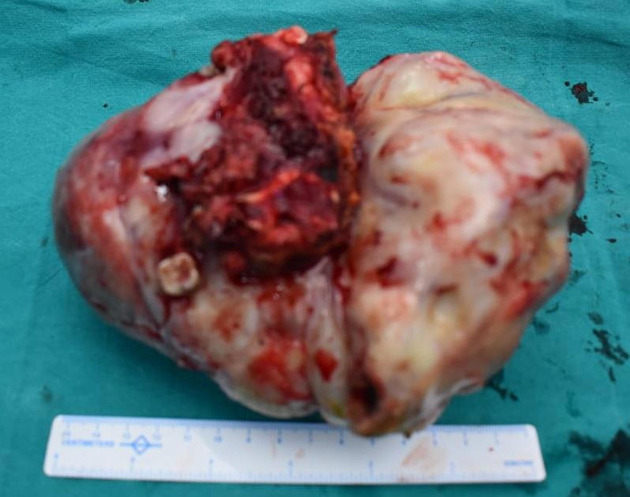
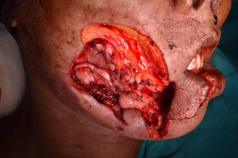
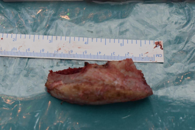
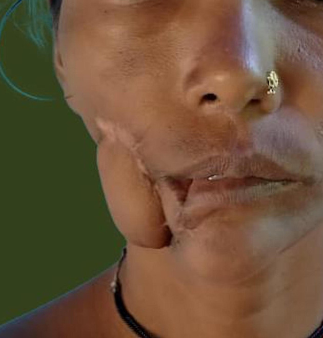
Psammomatoid Juvenile ossifying fibroma (PsJOF) is a rare benign fibro-osseous lesion characterised to grow to unusually large size very rapidly. Its usual presentation is in younger age group mostly children and predominately involving the Sino-Naso-Orbital region. Its aggressive nature gimmicks a malignant lesion but it is rather a benign lesion with a higher recurrence rate than the conventional ossifying fibroma but lacking metastatic potential. The high recurrence rate makes it essential that the lesion is not reconstructed immediately and thorough monitoring in the follow-up period. Lesion of such clinical importance needs to be diagnosed preoperatively to provide a better and radical surgical treatment option, but the variability in its presentation as seen in this case makes it even harder to diagnose. We aim to draw attention to the rare phenomena that PsJOF presents to help readers broaden their purview in diagnosis and thereby manage them accordingly.
Keywords: head and neck cancer; oral and maxillofacial surgery; otolaryngology / ENT.
© BMJ Publishing Group Limited 2021. No commercial re-use. See rights and permissions. Published by BMJ.
Conflict of interest statement
Competing interests: None declared.
Figures
Similar articles
-
Spheno-orbital juvenile psammomatoid ossifying fibroma: a case report and literature review.Childs Nerv Syst. 2021 Oct;37(10):3251-3255. doi: 10.1007/s00381-020-05004-8. Epub 2021 Jan 6. Childs Nerv Syst. 2021. PMID: 33404728 Review.
-
Juvenile Ossifying Fibroma of Maxilla.Kathmandu Univ Med J (KUMJ). 2018 Jul-Sept.;16(63):263-265. Kathmandu Univ Med J (KUMJ). 2018. PMID: 31719318
-
Psammomatoid juvenile ossifying fibroma: an analysis of 2 cases affecting the mandible with review of the literature.Oral Surg Oral Med Oral Pathol Oral Radiol. 2012 Jun;113(6):e40-5. doi: 10.1016/j.oooo.2011.08.005. Epub 2012 Feb 28. Oral Surg Oral Med Oral Pathol Oral Radiol. 2012. PMID: 22668716 Review.
-
Cemento-ossifying fibroma transforming to osteosarcoma.BMJ Case Rep. 2024 Jan 16;17(1):e257104. doi: 10.1136/bcr-2023-257104. BMJ Case Rep. 2024. PMID: 38233000
-
Psammomatoid variant of juvenile ossifying fibroma.Indian J Pathol Microbiol. 2018 Jul-Sep;61(3):443-445. doi: 10.4103/IJPM.IJPM_577_17. Indian J Pathol Microbiol. 2018. PMID: 30004078
References
Publication types
MeSH terms
LinkOut - more resources
Full Text Sources
Other Literature Sources
Medical
Research Materials
Miscellaneous