Review
. 2021 Apr;70(4):823-830.
doi: 10.2337/db20-1111.

Pregnancies in Diabetes and Obesity: The Capacity-Load Model of Placental Adaptation

Affiliations
Review

Pregnancies in Diabetes and Obesity: The Capacity-Load Model of Placental Adaptation

Gernot Desoye et al. Diabetes. 2021 Apr.

Abstract

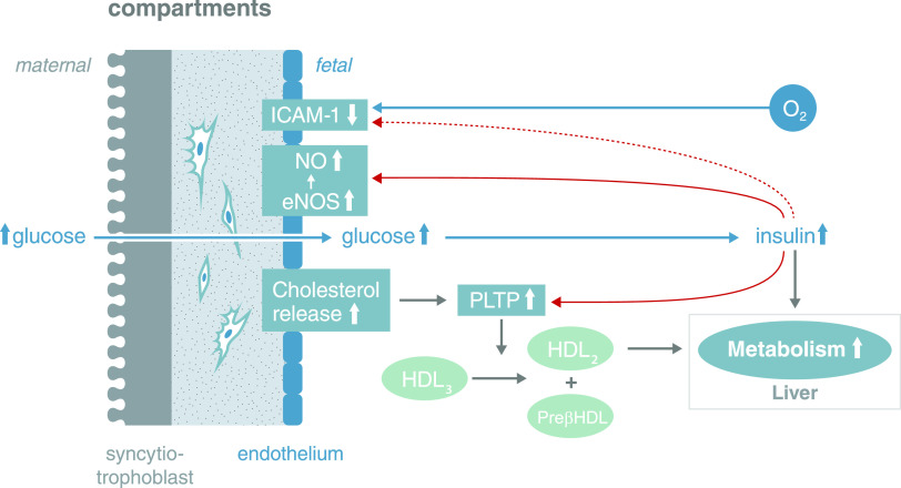
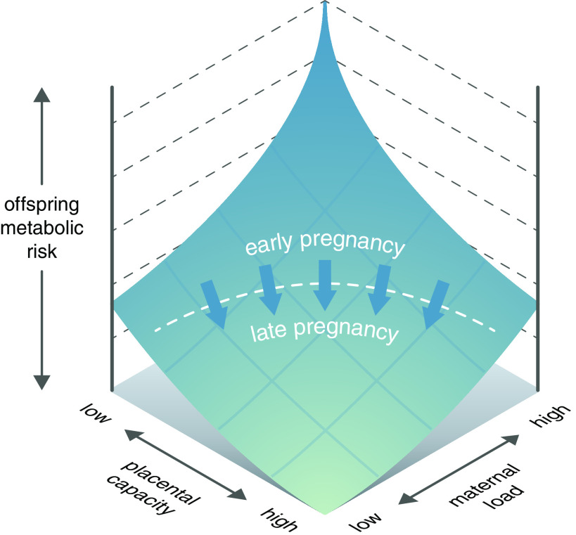
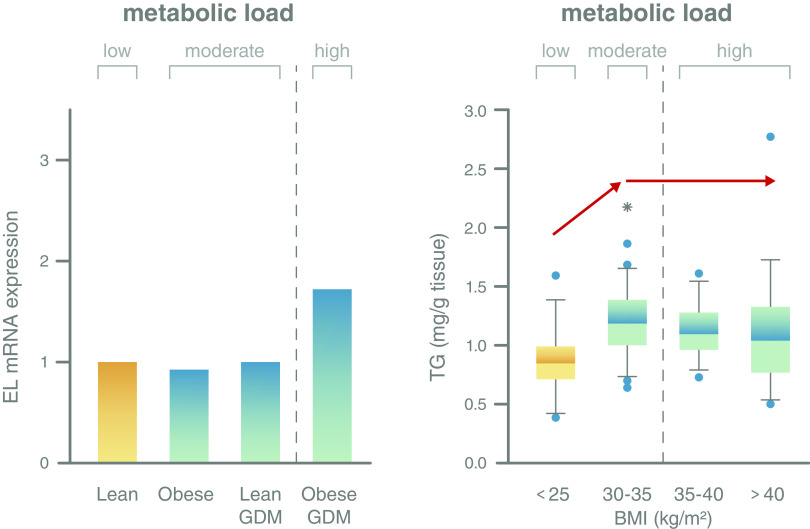
Excess nutritional supply to the growing fetus, resulting from maternal diabetes and obesity, is associated with increased risks of fetal maldevelopment and adverse metabolic conditions in postnatal life. The placenta, interposed between mother and fetus, serves as the gateway between the two circulations and is usually considered to mediate maternal exposures to the fetus through a direct supply line. In this Perspective, however, we argue that the placenta is not an innocent bystander and mounts responses to fetal "signals of distress" to sustain its own adequate function and protect the fetus. We describe several types of protection that the placenta can offer the fetus against maternal metabolic perturbations and offer a theoretical model of how the placenta responds to the intrauterine environment in maternal diabetes and obesity to stabilize the fetal environment. Our approach supports growing calls for early screening and control of pregnancy metabolism to minimize harmful fetal outcomes.


Figures

Figure 1
Figure 1
Fetal signals related to excess nutritional supply facilitate placental adaptation to prevent atherosclerotic plaques being formed. Fetoplacental endothelial cells synthesize more cholesterol in GDM than in normal pregnancies (19). At the same time, two cholesterol efflux transporters (ABCA1, ABCG1) are upregulated through activation of the LXR transcription factor in response to higher concentrations of circulating and intraendothelial oxysterols formed by ROS-induced cholesterol oxidation. Cholesterol efflux from these endothelial cells is also enhanced by insulin-induced upregulation of phospholipid transfer protein (PLTP) on the surface of the endothelial cells in GDM (20,72). This enzyme transfers cholesterol from HDL3 to HDL2, while pre-β HDL remains. Pre-β HDL can pick up cholesterol from the endothelial cells, as it is a cholesterol acceptor (73). The majority of HDL2 will be taken up by the fetal liver and the cholesterol converted into bile acids. Thus, there is a very efficient system for removing free cholesterol from fetoplacental endothelial cells and the fetoplacental circulation to avoid fetal hypercholesterolemia under conditions of GDM. A second system to prevent formation of atherosclerotic plaques involves the downregulation of intercellular adhesion molecule 1 (ICAM-1) expressed on the surface of fetoplacental endothelial cells in GDM (74). ICAM-1 mediates the adhesion of leukocytes to endothelial cells, which then transmigrate into the subendothelial space. Outside pregnancy, this mechanism plays a pivotal role in the inflammatory component of atherosclerosis (75). Loss of endothelial cell surface and soluble ICAM-1 in GDM may be induced, among other factors, by fetal insulin (76). Fetal insulin also increases endothelial nitric oxide synthase (eNOS) and nitric oxide (NO) synthesis (12) providing atheroprotection (77). These three systems, and probably more, act in concert to ultimately protect the fetoplacental circulation. Note: This is a schematic depicting the coordinated action of the players and not their precise location. PLTP is located on the endothelial surface.
Figure 2
Figure 2
Placental homeostatic capacity/efficiency model. During pregnancy, the placenta has a homeostatic capacity that will maintain fetoplacental homeostasis and determine the efficiency, with which the placenta protects the fetus from adverse consequences of a disturbed intrauterine environment. Up to a certain level of maternal metabolic load, the placenta can respond to signals such as insulin and orchestrate adaptive homeostatic responses that preserve an optimal metabolic milieu for fetal development. We hypothesize that this capacity is negligible during the early weeks in pregnancy and fully developed at the end. However, above the limiting threshold, such placenta capacity is exhausted and adverse fetal effects ensue. This threshold may differ according to both maternal and fetal traits (53).
Figure 3
Figure 3
Placental homeostatic capacity has its limits resulting in potentially adverse changes when the metabolic load exceeds this limit. Left panel: Expression of endothelial lipase (EL), a gene that is involved in lipid metabolism and perhaps fatty acid supply to the fetus, is stable in maternal obesity and GDM and only responds when metabolic load is high, i.e., in GDM and obesity (64). Importantly, the association of maternal obesity with adverse outcomes such as high neonatal fat mass is independent of maternal (gestational) diabetes (5). Right panel: Placental accumulation of triglycerides (TG) as a means to protect against lipotoxicity (63) increases with increasing maternal metabolic load, i.e., between lean women and women with class I obesity (BMI 30–35 kg/m2). This capacity is exhausted with a further increase in load, i.e., in women with class II and III obesity (BMI 35–40 and >40 kg/m2), and no further additional triglycerides are stored (64).

References

    1. Myatt L. Placental adaptive responses and fetal programming. J Physiol 2006;572:25–30 - PMC - PubMed
    1. Gauster M, Desoye G, Tötsch M, Hiden U. The placenta and gestational diabetes mellitus. Curr Diab Rep 2012;12:16–23 - PubMed
    1. Hirschmugl B, Crozier S, Matthews N, et al. Relation of placental alkaline phosphatase expression in human term placenta with maternal and offspring fat mass. Int J Obes 2018;42:1202–1210 - PMC - PubMed
    1. Catalano PM, Thomas A, Huston-Presley L, Amini SB. Phenotype of infants of mothers with gestational diabetes. Diabetes Care 2007;30(Suppl. 2):S156–S160 - PubMed
    1. McIntyre HD, Catalano P, Zhang C, Desoye G, Mathiesen ER, Damm P. Gestational diabetes mellitus. Nat Rev Dis Primers 2019;5:47. - PubMed