. 2021 Sep;11(9):2200-2215.
doi: 10.1158/2159-8290.CD-20-1066. Epub 2021 Mar 19.

ZFTA-RELA Dictates Oncogenic Transcriptional Programs to Drive Aggressive Supratentorial Ependymoma

Affiliations

ZFTA-RELA Dictates Oncogenic Transcriptional Programs to Drive Aggressive Supratentorial Ependymoma

Amir Arabzade et al. Cancer Discov. 2021 Sep.

Abstract

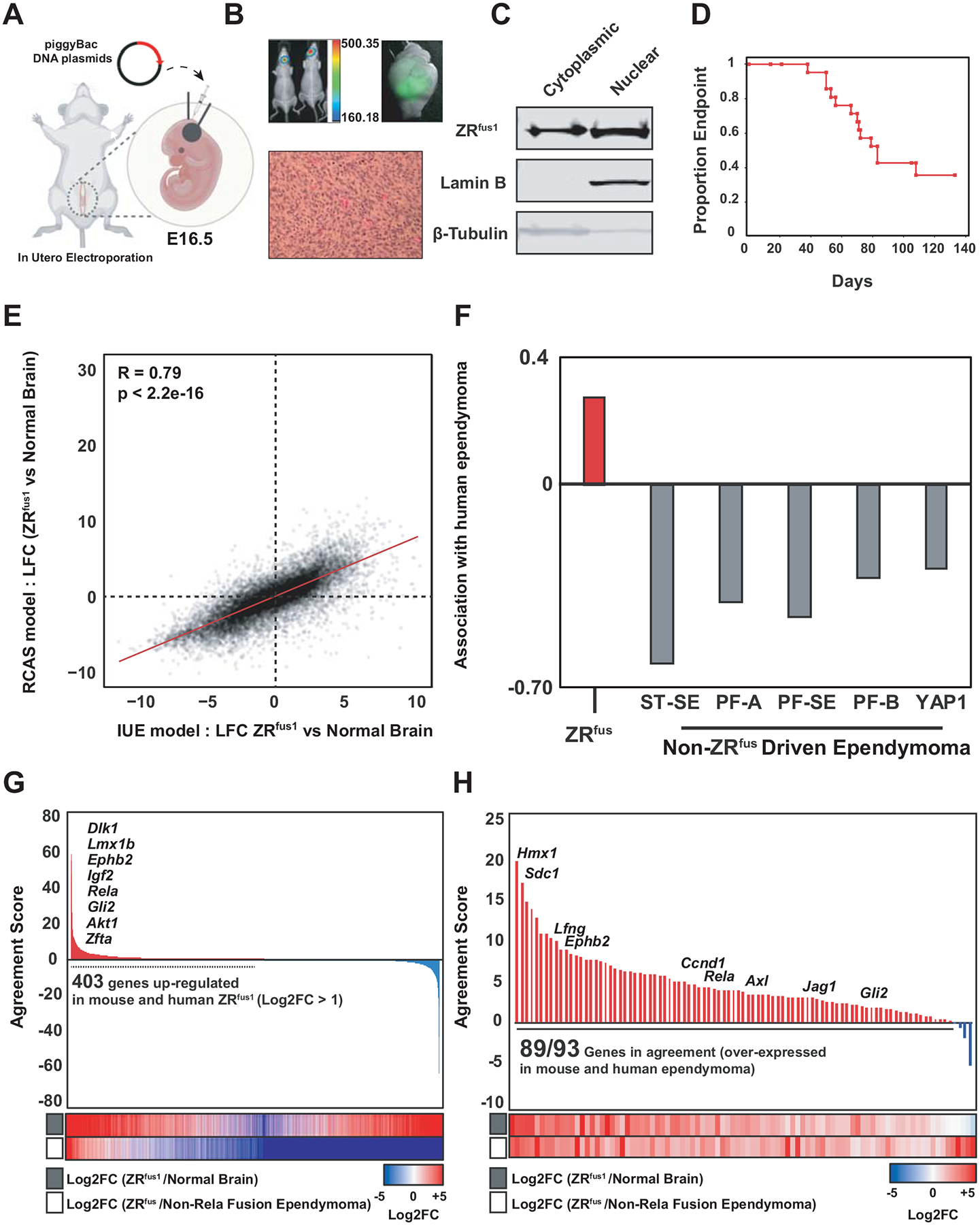
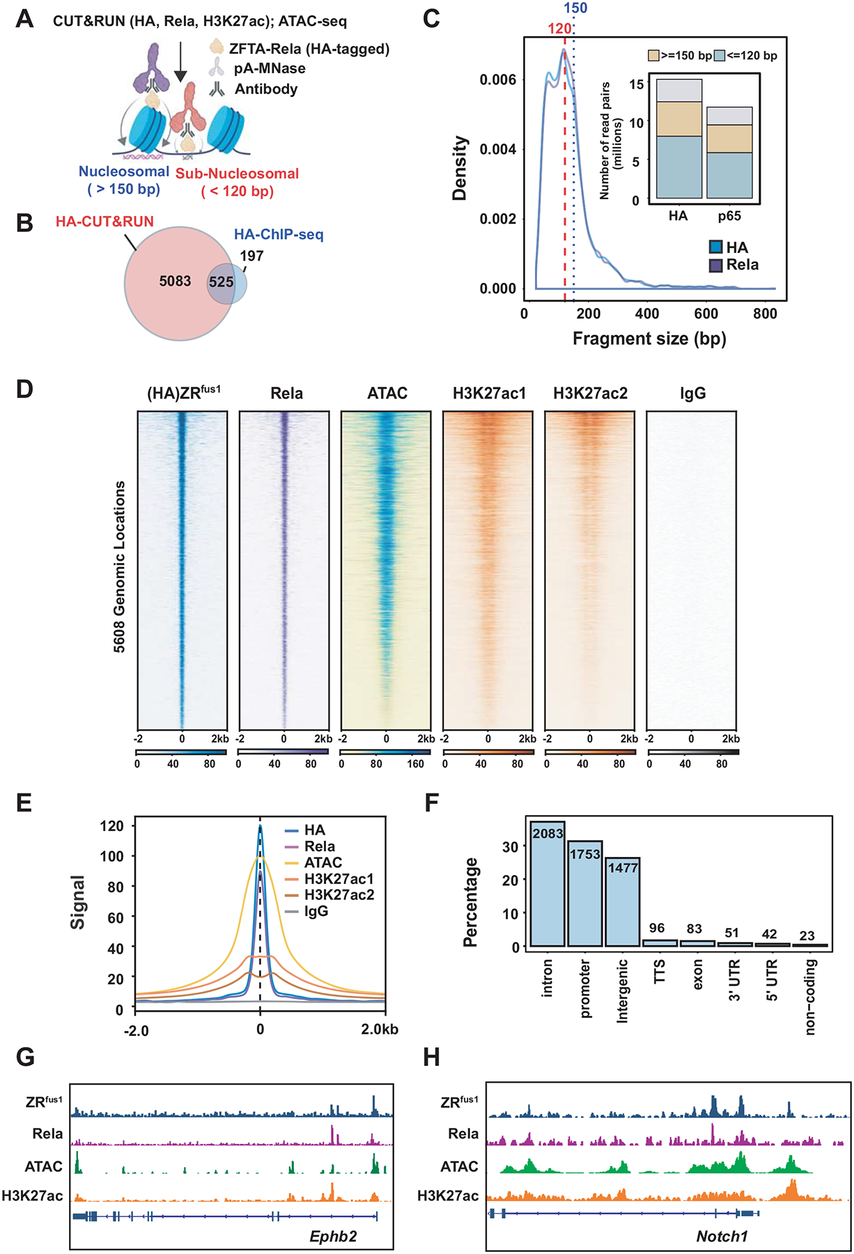
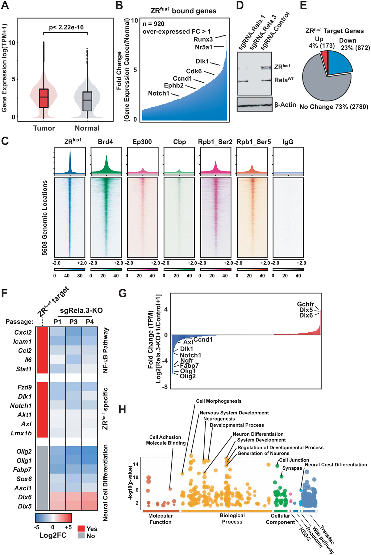
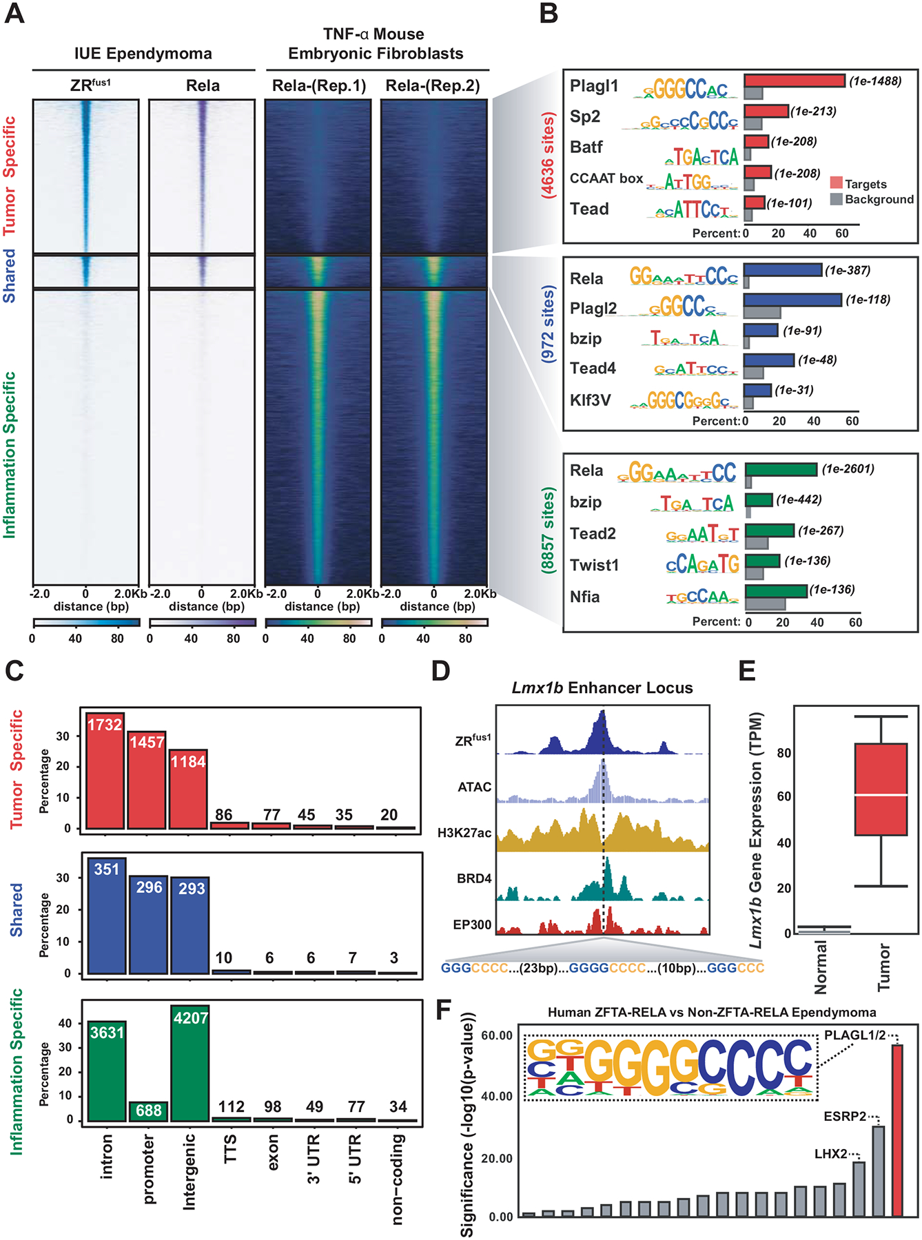
More than 60% of supratentorial ependymomas harbor a ZFTA-RELA (ZRfus) gene fusion (formerly C11orf95-RELA). To study the biology of ZRfus, we developed an autochthonous mouse tumor model using in utero electroporation (IUE) of the embryonic mouse brain. Integrative epigenomic and transcriptomic mapping was performed on IUE-driven ZRfus tumors by CUT&RUN, chromatin immunoprecipitation sequencing, assay for transposase-accessible chromatin sequencing, and RNA sequencing and compared with human ZRfus-driven ependymoma. In addition to direct canonical NFκB pathway activation, ZRfus dictates a neoplastic transcriptional program and binds to thousands of unique sites across the genome that are enriched with PLAGL family transcription factor (TF) motifs. ZRfus activates gene expression programs through recruitment of transcriptional coactivators (Brd4, Ep300, Cbp, Pol2) that are amenable to pharmacologic inhibition. Downstream ZRfus target genes converge on developmental programs marked by PLAGL TF proteins, and activate neoplastic programs enriched in Mapk, focal adhesion, and gene imprinting networks. SIGNIFICANCE: Ependymomas are aggressive brain tumors. Although drivers of supratentorial ependymoma (ZFTA- and YAP1-associated gene fusions) have been discovered, their functions remain unclear. Our study investigates the biology of ZFTA-RELA-driven ependymoma, specifically mechanisms of transcriptional deregulation and direct downstream gene networks that may be leveraged for potential therapeutic testing.This article is highlighted in the In This Issue feature, p. 2113.


Conflict of interest statement

Conflicts of Interest:

The authors declare no potential conflicts of interest

Figures

Figure 1
Figure 1. Establishment of a native ZFTA-Rela driven mouse model by embryonic in utero electroporation (IUE).
(A) Schematic of in utero electroporation technique used for introduction of piggyBAC DNA plasmids at embryonic day 16.5 (E16.5) (B) Luciferase, GFP imaging, and histology of IUE ZRfus1 murine ependymoma, and (C) confirmation of fusion protein nuclear localization. (D) Survival of mice reaching tumor endpoint as a result of ZRfus1expression. (E) Pearson correlation of IUE vs RCAS driven ZFTA-RELA mouse ependymoma(8) and (F) association with human ZFTA-RELA human tumor expression (18). (G) Comparison between genes upregulated in mouse IUE- ZRfus1 tumors vs matched normal brain and upregulated in ZRfus human ependymoma vs other ependymoma subtypes. Shared genes were restricted to those with a log2 fold change greater than 1. Agreement score represents the product of the fold change in mouse and human comparisons. (H) Waterfall plot of 93 ZFTA-Rela signature genes ordered by agreement score representing the product of Log2 fold change (ZRfus1/Normal Brain) and Log2 fold change (ZFTA-Rela/Non-Rela ependymoma).
Figure 2
Figure 2. ZFTA-Rela binds open and active chromatin of promoter and enhancer loci
(A) Schematic of proteins and epigenetic marks profiled using CUT&RUN, ChIP-seq, or ATAC-seq in IUE ZRfus1 ependymoma. (B) Venn diagram of the intersection between ZRfus1 peaks detected using CUT&RUN and ChIP-seq data. (C) CUT&RUN fragment size profiles of HA and Rela (p65). (D-E) Localization of ZRfus1 by HA-CUT&RUN and Rela-CUT&RUN in open chromatin (ATAC-seq) and actively transcribed chromatin (H3K27ac CUT&RUN). (F) Distribution of ZRfus1 binding sites at different genomic regions. (G-H) Binding profiles of ZRfus1 at the promoters and enhancers of ependymoma oncogenes.
Figure 3
Figure 3. Recruitment of transcriptional activation proteins to ZFTA-Rela binding sites
(A-B) Gene expression of ZRfus1 targets in mouse tumors versus contralateral normal brain (C) Recruitment of transcriptional co-regulators Brd4, Ep300, Crebbp, Ser2-Pol2, and Ser5-Pol2 to ZRfus1 binding sites as compared to IgG control. (D) CRISPR-CAS9 KO of ZRfus1 and Rela (E) Pie chart depicting the number of genes that were down-regulated, up-regulated, or exhibited no change in expression between non-targeting and Rela KO (F) Heatmap of selected genes in the NF-kB, ZRfus1 targets, and neuronal differentiation pathway that were down-regulated or up-regulated (G) Differential gene expression between non-targeting and Rela KO experiments across three cell passages. Represented in the waterfall plot is the Log2 fold-change between KO versus non targeting across n = 24,428 genes/transcripts. (H) Pathway analysis of the top down-regulated genes following Rela-KO as compared to non-targeting controls.
Figure 4
Figure 4. ZFTA-Rela engages NF-κB and Plagl (‘GGGCC’) motifs
(A) Comparison of HA- and Rela- CUT&RUN binding in ZRfus1 ependymoma against mouse embryonic fibroblasts cells treated with TNF-α. Motif enrichment analysis (B) and Distribution of peaks at different genomic regions (C) for tumor-specific, shared, and inflammation-specific binding sites. (D) Example of ZRfus1 binding at the Lmx1b locus, recruitment of Brd4 and Ep300, and presence of multiple Plagl motifs. (E) Lmx1b gene expression in IUE ZRfus1 tumor versus normal brain. (F) Comparison of DNA motifs identified in human ZFTA-RELA and non-ZFTA-RELA ependymoma using HOMER (see methods).
Figure 5
Figure 5. ZFTA-RELA disrupts core regulatory circuitry programs to drive SE gene expression
(A) Hockey-stick plot depicting super enhancers detected in IUE: ZRfus1 murine ependymoma (B) Venn diagram showing overlap of HA: ZRfus1 peaks and SEs (C) Box-violin plot depicting up-regulation of SE associated genes in tumor vs. normal brain (D) HA-ZRfus1 CUT&RUN, ATAC-seq, Brd4 CUT&RUN, and H3K27ac CUT&RUN shown at the Dlk1 locus (E) Dlk1 expression in mouse and human ZRfus ependymoma compared to normal brain and other ependymoma subtypes, respectively (F) Schematic of identification of core regulatory circuit (CRC) transcription factors (G) Top CRC module enriched in IUE-ZRfus1 ependymoma including predicted binding sites and engagement of ZRfus1 at Plagl1/2 and Rela motifs (H) CUT&RUN localization of Plagl2, HA-ZRfus1 and Rela at super enhancers that harbor the Plagl1/2 or Rela motif.
Figure 6
Figure 6. Pathway enrichment of ZFTA-RELA specific and inflammatory-shared target genes
(A) Tumor-specific programs enriched with Plagl TF motifs. Color of nodes highlight differential gene expression between mouse ZRfus1 and contralateral brain. (B) Integration of ZRfus1 bound and over-expressed genes with the Washington University Drug-Gene Interaction database to identify candidate drugs and small molecule inhibitors as potential therapeutic candidates against ependymoma.

References

    1. Merchant TE, Bendel AE, Sabin ND, Burger PC, Shaw DW, Chang E, et al.Conformal Radiation Therapy for Pediatric Ependymoma, Chemotherapy for Incompletely Resected Ependymoma, and Observation for Completely Resected, Supratentorial Ependymoma. J Clin Oncol 2019;37(12):974–83 doi 10.1200/jco.18.01765. - DOI - PMC - PubMed
    1. Pajtler KW, Wen J, Sill M, Lin T, Orisme W, Tang B, et al.Molecular heterogeneity and CXorf67 alterations in posterior fossa group A (PFA) ependymomas. Acta Neuropathol 2018;136(2):211–26 doi 10.1007/s00401-018-1877-0. - DOI - PMC - PubMed
    1. Pajtler KW, Witt H, Sill M, Jones DT, Hovestadt V, Kratochwil F, et al.Molecular Classification of Ependymal Tumors across All CNS Compartments, Histopathological Grades, and Age Groups. Cancer cell 2015;27(5):728–43 doi 10.1016/j.ccell.2015.04.002. - DOI - PMC - PubMed
    1. Hoffman LM, Donson AM, Nakachi I, Griesinger AM, Birks DK, Amani V, et al.Molecular subgroup-specific immunophenotypic changes are associated with outcome in recurrent posterior fossa ependymoma. Acta Neuropathol 2014;127(5):731–45 doi 10.1007/s00401-013-1212-8. - DOI - PMC - PubMed
    1. Bayliss J, Mukherjee P, Lu C, Jain SU, Chung C, Martinez D, et al.Lowered H3K27me3 and DNA hypomethylation define poorly prognostic pediatric posterior fossa ependymomas. Science translational medicine 2016;8(366):366ra161 doi 10.1126/scitranslmed.aah6904. - DOI - PMC - PubMed

Publication types