

Review
doi: 10.1097/SCS.0000000000007618.
Ocular Related Sports Injuries
Affiliations
- PMID: 33741878
- PMCID: PMC8192440
- DOI: 10.1097/SCS.0000000000007618
Item in Clipboard
Review
Ocular Related Sports Injuries
J Craniofac Surg.
.
Abstract
Ocular injuries occur frequently in sports, affecting the globe, surrounding soft tissues, and the orbital bony structure. This review provides the craniofacial surgeon a broad general overview of epidemiology, mechanism of disease, and prevention.
Copyright © 2021 by Mutaz B. Habal, MD.
Conflict of interest statement
The authors report no conflicts of interest.
Figures
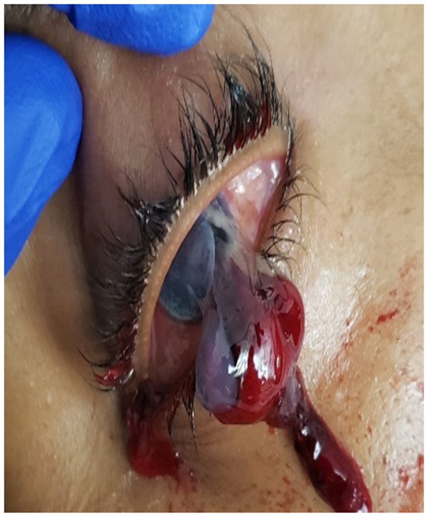
Intraocular contents have extruded onto the lower lid of a patient with a ruptured globe (full thickness break in the inferotemporal cornea). Photo courtesy of Benjamin Lin, MD.
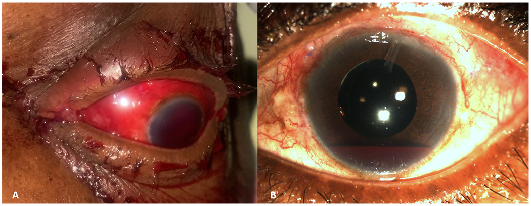
An 8 ball hyphema and diffuse subconjunctival hemorrhage is noted in this patient who presented after blunt trauma to the eye (A). Photo courtesy of Marissa Shoji, MD. A hyphema can be seen layering in the bottom of the anterior chamber of this patient with a history of previous cataract surgery and placement of a glaucoma drainage implant (B). Photo courtesy of Raquel Goldhardt, MD.
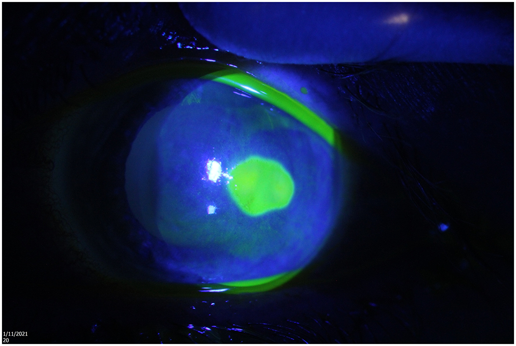
A corneal epithelial defect is fluorescing after cobalt blue light is being shone on an eye that has been stained with fluorescein. Photo courtesy of Alison Bozung, OD.
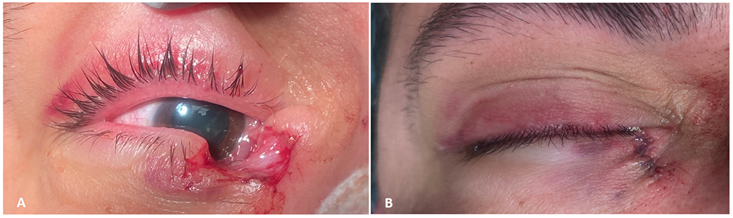
A full thickness lower lid laceration is demonstrated in this patient with a history of blunt trauma to the face (A) and after surgical repair (B). Photo courtesy of Marissa Shoji, MD.
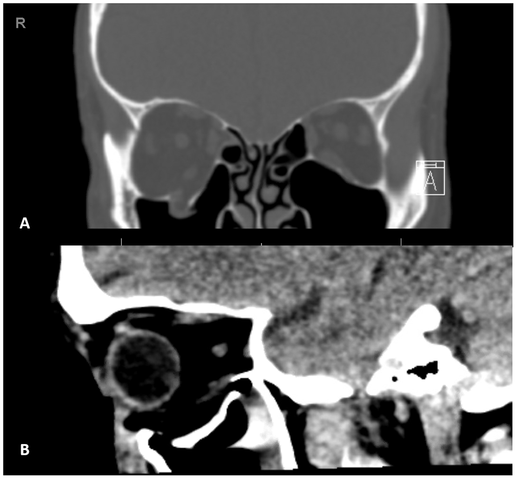
CT scan of a 14 Year old female who was struck in the left orbit by a softball, showing orbital wall fractures. (A) Coronal view: Left medial and floor fracture. Notice fracture propagation along the roof of the infra orbital canal and lamina papyracea. White arrow points to intra-orbital air. (B) Axial view better depicts the medial wall fracture. (C) Sagittal view allows better appreciation of the depressed orbital floor fracture.
Similar articles
-
Ocular injuries in hockey.Int Ophthalmol Clin. 1988 Fall;28(3):228-31. doi: 10.1097/00004397-198802830-00013. Int Ophthalmol Clin. 1988. PMID: 3403182 No abstract available.
-
Sports injuries: an ounce of prevention and a pound of cure.Eye Contact Lens. 2011 May;37(3):160-3. doi: 10.1097/ICL.0b013e31821790db. Eye Contact Lens. 2011. PMID: 21471814 Review.
-
Epidemiology of Sports-Related Eye Injuries in the United States.JAMA Ophthalmol. 2016 Dec 1;134(12):1382-1390. doi: 10.1001/jamaophthalmol.2016.4253. JAMA Ophthalmol. 2016. PMID: 27812702
-
Eye injuries in Canadian sports and recreation, 1972-2002.Can J Ophthalmol. 2002 Jun;37(4):253-5. doi: 10.1016/s0008-4182(02)80118-1. Can J Ophthalmol. 2002. PMID: 12095100 No abstract available.
-
The epidemiology of sports-related ocular trauma.Int Ophthalmol Clin. 1988 Fall;28(3):199-202. doi: 10.1097/00004397-198802830-00003. Int Ophthalmol Clin. 1988. PMID: 3042659 Review. No abstract available.
Cited by
-
An Analysis of Ocular Trauma Resulting From Pediatric Sports Injuries.Clin Ophthalmol. 2025 Feb 12;19:507-517. doi: 10.2147/OPTH.S493655. eCollection 2025. Clin Ophthalmol. 2025. PMID: 39963522 Free PMC article.
-
Discrepancy of eye injuries in mechanism, clinical features, and vision prognosis by different causative sports.Front Public Health. 2023 Oct 18;11:1182647. doi: 10.3389/fpubh.2023.1182647. eCollection 2023. Front Public Health. 2023. PMID: 37920581 Free PMC article.
-
Retinal Screening in High-Performance Athletes: A Retrospective Analysis of Asymptomatic Peripheral Lesions in Collision and Non-Collision Sports.Sports Med Open. 2025 Jun 11;11(1):74. doi: 10.1186/s40798-025-00869-y. Sports Med Open. 2025. PMID: 40498160 Free PMC article.
-
International Olympic Committee (IOC) consensus paper on sports-related ophthalmology issues in elite sports.BMJ Open Sport Exerc Med. 2023 Jul 19;9(3):e001644. doi: 10.1136/bmjsem-2023-001644. eCollection 2023. BMJ Open Sport Exerc Med. 2023. PMID: 37485004 Free PMC article.
-
Vitreoretinal Injury Associated with Sports Ball Ocular Trauma.Clin Ophthalmol. 2025 Jun 23;19:1931-1943. doi: 10.2147/OPTH.S507399. eCollection 2025. Clin Ophthalmol. 2025. PMID: 40584248 Free PMC article. Review.
References
-
- Haring RS, Sheffield ID, Canner JK, Schneider EB. Epidemiology of Sports-Related Eye Injuries in the United States. JAMA Ophthalmol. 2016;134(12):1382. - PubMed
-
- Miller KN, Collins CL, Chounthirath T, Smith GA. Pediatric Sports- and Recreation-Related Eye Injuries Treated in US Emergency Departments. Pediatrics. 2018;141(2). - PubMed
-
- Goldstein MH, Wee D. Sports Injuries: An Ounce of Prevention and a Pound of Cure. Eye Contact Lens Sci Clin Pract. 2011;37(3):160–163. - PubMed
-
- Toldi JP, Thomas JL. Evaluation and Management of Sports-Related Eye Injuries. Curr Sports Med Rep. 2020;19(1):29–34. - PubMed
-
- Cass SP. Ocular injuries in sports. Curr Sports Med Rep. 2012;11(1):11–15. - PubMed
Publication types
MeSH terms
Grants and funding
LinkOut - more resources
Full Text Sources
Other Literature Sources
Medical
