. 2021 Mar 19;12(1):1726.
doi: 10.1038/s41467-021-21972-0.

Alpha-1 antitrypsin inhibits TMPRSS2 protease activity and SARS-CoV-2 infection

Affiliations

Alpha-1 antitrypsin inhibits TMPRSS2 protease activity and SARS-CoV-2 infection

Lukas Wettstein et al. Nat Commun. .

Abstract

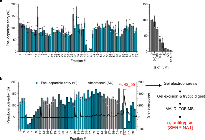
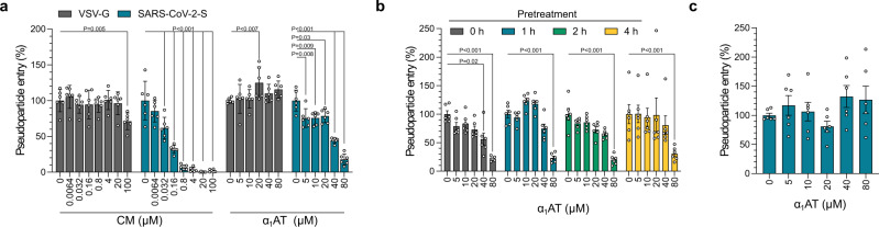
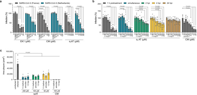
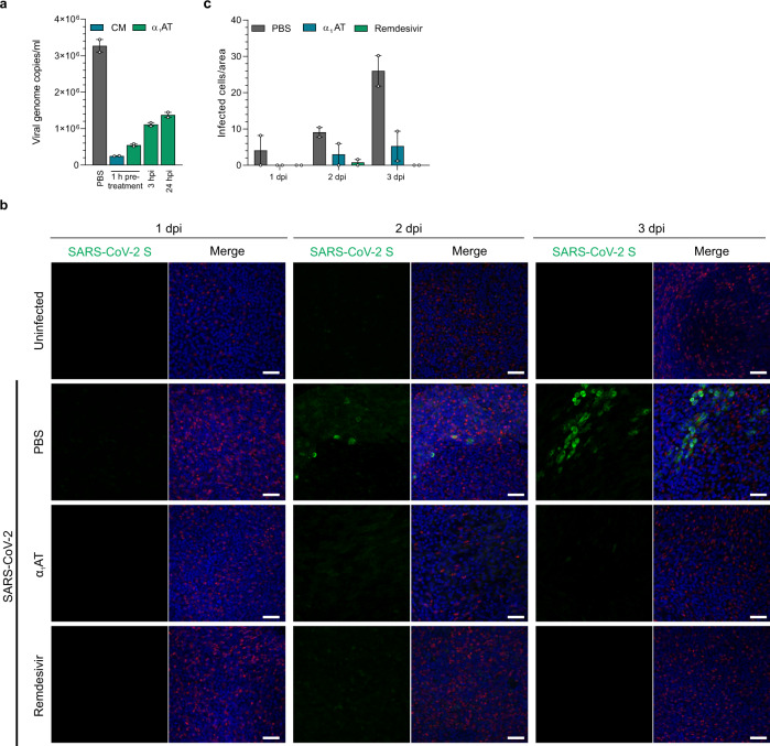
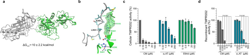
SARS-CoV-2 is a respiratory pathogen and primarily infects the airway epithelium. As our knowledge about innate immune factors of the respiratory tract against SARS-CoV-2 is limited, we generated and screened a peptide/protein library derived from bronchoalveolar lavage for inhibitors of SARS-CoV-2 spike-driven entry. Analysis of antiviral fractions revealed the presence of α1-antitrypsin (α1AT), a highly abundant circulating serine protease inhibitor. Here, we report that α1AT inhibits SARS-CoV-2 entry at physiological concentrations and suppresses viral replication in cell lines and primary cells including human airway epithelial cultures. We further demonstrate that α1AT binds and inactivates the serine protease TMPRSS2, which enzymatically primes the SARS-CoV-2 spike protein for membrane fusion. Thus, the acute phase protein α1AT is an inhibitor of TMPRSS2 and SARS-CoV-2 entry, and may play an important role in the innate immune defense against the novel coronavirus. Our findings suggest that repurposing of α1AT-containing drugs has prospects for the therapy of COVID-19.


Conflict of interest statement

The authors declare no competing interests.

Figures

Fig. 1
Fig. 1. Identification of α1AT as SARS-CoV-2 inhibitor.
a Caco2 cells were treated with peptide/protein containing fractions of the bronchoalveolar lavage library (or EK1 peptide as inhibitor control) and transduced with luciferase-encoding lentiviral SARS-CoV-2 spike pseudoparticles. b Caco2 cells were treated with subfractions of mother fraction 42 (see a) of the bronchoalveolar lavage library and transduced with lentiviral SARS-CoV-2 spike pseudoparticles. Blue columns represent pseudoparticle entry and black line absorbance at 280 nm of the corresponding fraction. Transduction rates in a and b were determined 2 days after the addition of pseudoparticles by measuring luciferase activities in cell lysates. The means ± SEM from n = 2 (a) or individual datapoints from n = 1 (b) independent experiments are shown, each performed in biological duplicates. The active fraction 42_55 (red box) was analyzed via gel electrophoresis, gel excision, tryptic digest, and MALDI-TOF-MS to identify α1AT, a serine protease inhibitor (serpin). Source data are provided as a Source data file.
Fig. 2
Fig. 2. α1AT inhibits SARS-CoV-2 spike but not VSV-G mediated pseudovirus entry.
a Prolastin (α1AT) and the control small molecule inhibitor camostat mesylate (CM) were added to Caco2 cells 1 h prior to transduction of cells with rhabdoviral SARS-CoV-2 spike (blue) or VSV-G pseudoparticles (gray). b Caco2 cells were treated for the indicated hours with Prolastin (α1AT) and were then transduced with rhabdoviral SARS-CoV-2 spike pseudoparticles. c Prolastin (α1AT) was added 2 h post transduction of Caco2 cells with rhabdoviral SARS-CoV-2 spike pseudoparticles. Transduction rates in ac were determined at 16 h after addition of pseudoparticles by measuring luciferase activities in cell lysates. The mean ± SEM from n = 2 experiments in biological triplicates are shown (2-way ANOVA with Dunett´s multiple comparison test). Source data are provided as a Source data file.
Fig. 3
Fig. 3. α1AT inhibits SARS-CoV-2 infection and replication.
a TMPRSS2-expressing Vero E6 cells were treated with Prolastin (α1AT), EK1 or camostat mesylate (CM) for 1 h, and infected with SARS-CoV-2 isolates either from France (gray) or the Netherlands (blue) at a MOI of 0.001. Virus-induced cytopathic effects were assessed at 2 days post infection by MTS assay (see Supplementary Fig. 4 a, b for raw and cell viability data). b TMPRSS2-expressing Vero E6 cells were treated with Prolastin (α1AT) at indicated timepoints prior to, simultaneously with or post infection with SARS-CoV-2 at a MOI of 0.001. Camostat mesylate (CM) control was added 1 h prior to infection. Infection rates were assessed at 2 days post infection by MTS assay (see also Supplementary Fig. 5a, b for raw and cell viability data). c TMPRSS2-expressing Vero E6 cells were treated with Prolastin (α1AT) or CM 1 h prior to, simultaneously with or 1.5 h post infection with SARS-CoV-2. At 1.5 h post infection cellulose overlay was performed. At 2 days post infection, cells were stained with crystal violet (see Supplementary Fig. 6a, b) and plaque areas were quantified. The mean ± SEM from n = 1 (a, EK1 and CM) or n = 2 independent experiments in biological triplicates (a, α1AT, b) or duplicates (c) and quadruplicates (c, infected) are shown. (2-way ANOVA with Dunett´s multiple comparison test (a, b), ordinary one-way ANOVA with Dunett´s multiple comparison test (c)). Source data are provided as a Source data file.
Fig. 4
Fig. 4. α1AT inhibits SARS-CoV-2 replication in primary human airway cells.
a Small airway epithelial cells (SAECs) were treated with 100 µM of Prolastin (α1AT, green) 1 h prior to, or 3 and 24 h post infection (hpi) with SARS-CoV-2 at a MOI of 1. 100 µM CM (blue) added 1 h prior to infection served as control. Immediately after the inoculum was removed (0 dpi) and at day 6 post infection, supernatants were harvested and subjected to RT-qPCR specific for SARS-CoV-2 ORF1b nsp14. Virus titers from day 0 were subtracted from titers at day 6. The means of technical duplicates of one experiment performed in triplicates are shown. b The apical and basal site of human airway epithelial cells (HAEC) grown at air–liquid interface was exposed to PBS, α1AT (10 µM or 0.5 mg/ml) and remdesivir (5 µM) and then inoculated with SARS-CoV-2 (9.25 × 102 PFU) for 2 h. Cells were fixed at day 1, 2, and 3 post infection, stained with DAPI (cell nuclei, blue), a SARS-CoV-2 specific spike antibody (SARS-CoV-2 S, green) and an α-tubulin-specific antibody (red). Images shown are derived from one donor and represent maximum projections of serial sections along the basolateral to apical cell axis. Scale bar: 50 μm. c Number of infected cells per area in mock- (PBS, gray), α1AT- (blue) or remdesivir-treated (green), SARS-CoV-2 infected HAECs. Values represent the mean number of infected HAECs from 2 donors at 5 random spots per culture, treatment and day ± SEM. Source data are provided as a Source Data file. For more images, see Supplementary Fig. 7.
Fig. 5
Fig. 5. α1AT binds the extracellular region of TMPRSS2 and inhibits TMPRSS2 protease activity.
a Protein–protein docking analysis of a homology model for the TMPRSS2 extracellular fragment (green, PDB 1z8g) and α1AT (gray, PDB 3cwm) and computationally calculated binding free energy (ΔGcalc) of the complex. b Detailed view on α1AT-TMPRSS2 binding interface. The sidechains of α1AT (gray) residues are represented with sticks, while sidechains of TMPRSS2 (green) are shown with balls and sticks. Hydrogen atoms are omitted for clarity, carbon, oxygen, nitrogen or sulfur atoms of amino acid side chains depicted in light blue, red, dark blue or yellow, respectively. The hydrophobic patch near the TMPRSS2 catalytic triad is highlighted with a green transparent surface. c α1AT inhibits cell-associated TMPRSS2 activity. HEK293T cells were transfected with a TMPRSS2 expression plasmid and treated with Prolastin (α1AT, blue), camostat mesylate (CM, gray) or E-64d (green) followed by incubation with the fluorogenic TMRPSS2 protease substrate BOC-Gln-Ala-Arg-AMC. Graph shows the relative area under the curve analysis of fluorescence intensities over 2 h that were corrected by values for mock-transfected HEK293T cells. d α1AT inhibits recombinant TMPRSS2 enzyme activity. Recombinant human TMPRSS2 was mixed with Prolastin (α1AT, blue) or CM (gray) prior to addition of fluorogenic TMPRSS2 protease substrate BOC-Gln-Ala-Arg-AMC, graph shows relative fluorescence intensities after 3 h of incubation. The mean ± SEM of n = 2 (c) or n = 3 (d) independent experiments in biological duplicates are shown (ordinary one-way ANOVA with Dunett´s multiple comparison test). Source data are provided as a Source data file.

Similar articles

Cited by

References

    1. Hou YJ, et al. SARS-CoV-2 reverse genetics reveals a variable infection gradient in the respiratory tract. Cell. 2020;182:e14. doi: 10.1016/j.cell.2020.05.042. - DOI - PMC - PubMed
    1. Tay MZ, Poh CM, Rénia L, MacAry PA, Ng LFP. The trinity of COVID-19: immunity, inflammation and intervention. Nat. Rev. Immunol. 2020;20:363–374. doi: 10.1038/s41577-020-0311-8. - DOI - PMC - PubMed
    1. Gupta A, et al. Extrapulmonary manifestations of COVID-19. Nat. Med. 2020;26:1017–1032. doi: 10.1038/s41591-020-0968-3. - DOI - PMC - PubMed
    1. Vareille M, Kieninger E, Edwards MR, Regamey N. The airway epithelium: soldier in the fight against respiratory viruses. Clin. Microbiol. Rev. 2011;24:210–229. doi: 10.1128/CMR.00014-10. - DOI - PMC - PubMed
    1. Rogan MP, et al. Antimicrobial proteins and polypeptides in pulmonary innate defence. Respir. Res. 2006;7:1–11. doi: 10.1186/1465-9921-7-29. - DOI - PMC - PubMed

Publication types

MeSH terms